It’s time for another roundup of oral pathology cases to help you sharpen your diagnostic skills. Here are four cases from your colleagues that you can study, save, and use as a reference as you diagnose future cases from your clinical exams.
If you have recently encountered a pathology case in your practice, we encourage you to share it with us. One of the best sources of learning is peer-to-peer. Email your details to Bethany Montoya, editorial director of Clinical Insights newsletter, at [email protected]. We look forward to featuring you!
Stay tuned for more pathology content from DentistryIQ.
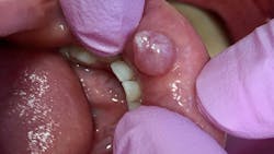
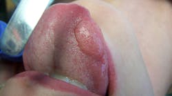
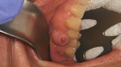
Patient: 6-year-old male
- 7 mm-plus soft, exophytic, dome-like lesion
- Normal lip color
- Fluid-filled with a slightly red base
- No pain, but patient was having a hard time eating due to position and size of lesion
- Patient had habit of biting his lip
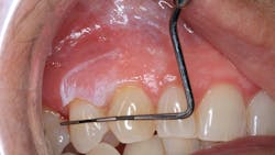
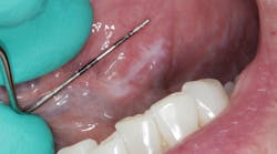
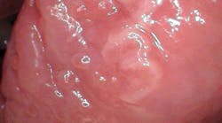
Patient: 66-year-old female
- 9 mm x 6 mm white, corrugated, irregular-bordered lesion
- Lesion located in upper right vestibular area
- Not tender to touch or palpation
- Not able to be removed or scraped off
- Large leukoplakic patch over erythematous tissue
Two weeks later …
- Lesion extended back toward first molar to mucolabial fold area
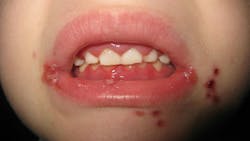
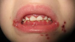
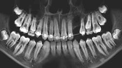
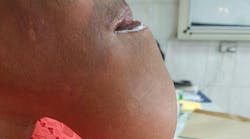
Patient: 2-year-old male
- Lump on right side of neck
- Ulcer-like lesions on buccal oral mucosa
- PCP diagnosed patient with hand, foot and mouth disease
A week later …
- Fire-engine red gingival tissue
- Manifestation of numerous perioral lesions
- Fever, loss of appetite, myalgia
- No lesions on hands or feet
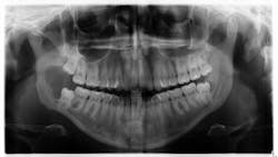
Patient: 22-year-old female
- Patient referred to oral surgeon for removal of wisdom teeth
- Following that, all contact with patient was lost
Seven years later …
- Large radiolucent lesion
- Significant bone destruction in mandible
- Inflamed tissue
- Tender to palpation
Dangerous lesion if left untreated
Editor’s note: This article first appeared in Clinical Insights newsletter, a publication of the Endeavor Business Media Dental Group. Read more articles and subscribe.
About the Author
Vicki Cheeseman
Associate Editor
Vicki Cheeseman is an associate editor in Endeavor Business Media’s Dental Group. She edits for Dental Economics, RDH, DentistryIQ, and Perio-Implant Advisory. She has a BS in mathematics and a minor in computer science. Early on she traded numbers for words and has been happy ever since. Vicki began her career with Dental Economics in 1987 and has been fascinated with how much media production has changed through the years, yet editorial integrity remains the goal. In her spare time, you’ll find her curled up with a book—editor by day, reader always.
