Oral pathology that you see during clinical exams is an excellent way to keep your detection skills sharp. One way to improve your clinical knowledge is to learn from cases your colleagues have seen.
Have you recently encountered a pathology case in your practice that you’d like to share? Email your details to Bethany Montoya, BAS, RDH, the editorial director of Clinical Insights. We look forward to featuring you!
Here are four pathology cases we have on file that you can use as a reference to help determine treatment in your future cases.

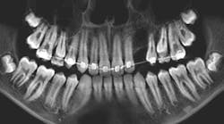
Patient: 37-year-old male
- Badly decayed teeth on the lower right side
- Apical radiolucency and associated swelling in the buccal vestibule
- Well-defined radiolucency above the apexes of teeth nos. 5-9
- Some expansion around the bone
- Lesion not fluctuant or mobile

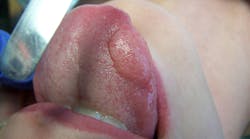
Patient: 50-year-old female
- Raised tissue mass between teeth nos. 4 and 5
- Penicillin allergy and history of type 1 and 2 herpes
- 6 mm pink, raised, firm mass of osseous tissue measuring 12x12 mm
- Lesion not tender to palpation and does not bleed easily when manipulated
“My doctor said I have an infection on one of my top right teeth”

Patient: 64-year-old female
- Red, inflamed tissue throughout the oral cavity
- Large, ulcerlike lesions on the right and left lateral borders of the tongue
- Well-defined edges with a slightly concave red center that is tender to palpation
- Vestibular tissue sloughs and hemorrhages easily
- Angular cheilitis observed

Patient: 66-year-old male
- Large radiolucency in the left mid-body of the mandible
- No oral lesions or vestibular swelling
- No extraoral visual or palpable swelling in the area
- No cervical lymphadenopathy
Read more pathology cases …

Editor’s note: This article first appeared in Clinical Insights newsletter, a publication of the Endeavor Business Media Dental Group. Read more articles and subscribe.