
Oral pathology that you see during clinical exams keeps your detection skills sharp. One way to improve your clinical knowledge is to learn from cases your colleagues have seen. Have you recently encountered a pathology case in your practice that you’d like to share? Email your details to Bethany Montoya, BAS, RDH, the editorial director of Clinical Insights. We look forward to featuring you!
Here are four pathology cases we have on file that you can use as a reference to help determine treatment in your future cases.

Patient: 62-year-old female
- 9 mm x 3 mm white leukoplakic patch of tissue on left ventral surface of the tongue
- Patient was unaware of its presence
- Not tender to palpation and could not be scraped off
- Noncontributory health history (patient was taking vitamin D supplements)

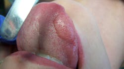
Patient: 21-year-old female
- 5 cm slow-growing, painless “lump” under the left ventral portion of the tongue
- Asymptomatic and no other lesions present
- No cervical lymphadenopathy
- Patient said the lump “popped” one time but came back a few weeks later

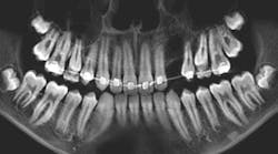
Patient: 7-year-old male
- Radiolucency appeared to focalize around the coronal portion of no. 7
- Ill-defined radiopacity within the osseous tissue under the area where primary tooth D would be located
- Not tender to palpation and no expansion in the vestibular area
- Patient had experienced trauma to the area a few years back while playing

Patient: 6-year-old male
- Tooth no. 9 had erupted normally after exfoliation of tooth F, but tooth E was still present
- Chief concern: Permanent teeth in the areas of nos. 7 and 8 might not be developing.
- Large radiodense area in the region apical to teeth D and E
- Otherwise healthy patient
Read more pathology cases …
Editor’s note: This article first appeared in Clinical Insights newsletter, a publication of the Endeavor Business Media Dental Group. Read more articles and subscribe.


Happy Laptop © Ocusfocus | Dreamstime.com

UnitoneVector / 1142658579 / iStock / Getty Images Plus
