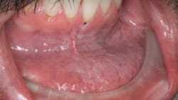
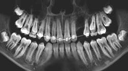
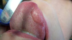
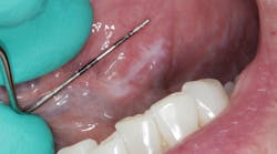
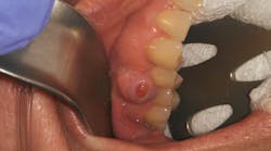
Any pathology that you see during oral exams can help you sharpen your clinical skills. One way to improve your clinical knowledge is to learn from cases your colleagues have seen.
If you have recently encountered a pathology case in your practice, please share it with us. Email your details to Bethany Montoya, BAS, RDH, the editorial director of Clinical Insights. We look forward to featuring you!
Here are four pathology cases from our files that you can use as a reference to help determine treatment in future cases.
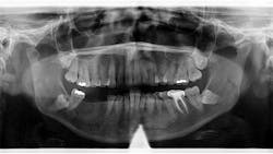
Patient: 50-year-old female
- 3 cm x 1.5 cm almost-vague radiodense lesion on lower left side around the apices of nos. 20-22
- Nonpalpable lesion
- Area not tender to palpation
- Patient taking prescription medication for an infection
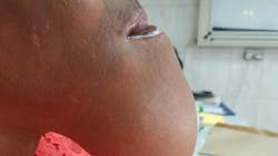
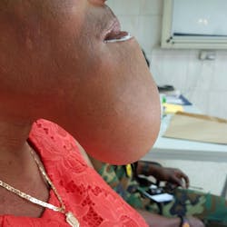
Patient: 40-year-old female
- Recurrent anterior mandibular swelling
- Two prior surgeries over the past six years to correct the condition
- Lesion previously removed when it was small and asymptomatic
- Patient pursued herbal treatment after second surgery
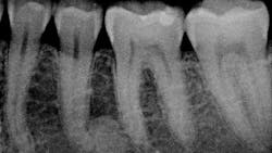
Patient: 41-year-old female
- Large, radiopaque mass just distal to apical third of no. 20
- Area not tender to palpation
- Tooth tests vital and WNL
- Normal health history
Patient: 27-year-old male
- Chief complaint: upper right tooth, third from the back, was bothering the patient progressively over the last few months
- Patient has slightly elevated blood pressure and is a chronic tobacco user
- Rough, corrugated-cardboard-like tissue in large area of lower left vestibule
- Patient is aware of the risks associated with tobacco
Editor’s note: This article first appeared in Clinical Insights newsletter, a publication of the Endeavor Business Media Dental Group. Read more articles and subscribe.
Read more pathology cases ...
About the Author
Vicki Cheeseman
Associate Editor
Vicki Cheeseman is an associate editor in Endeavor Business Media’s Dental Group. She edits for Dental Economics, RDH, DentistryIQ, and Perio-Implant Advisory. She has a BS in mathematics and a minor in computer science. Early on she traded numbers for words and has been happy ever since. Vicki began her career with Dental Economics in 1987 and has been fascinated with how much media production has changed through the years, yet editorial integrity remains the goal. In her spare time, you’ll find her curled up with a book—editor by day, reader always.
