Any pathology you note during your clinical exams can help sharpen your detection skills. A great way to improve your clinical knowledge is to learn from cases your colleagues have seen.
If you have recently encountered a pathology case in your practice, please share it with us. Email your details to Bethany Montoya, BAS, RDH, the editorial director of Clinical Insights. We look forward to featuring you!
Here are four pathology cases from our files that you can use as a reference to help determine treatment in future cases.
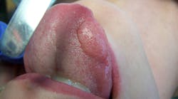
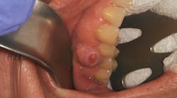
Patient: 8-year-old female
- 6 mm x 6 mm raised, round soft-tissue lesion
- Red outside border and a white center
- Not tender to palpation
- Located under the left side of the tongue along the sublingual salivary gland ducts
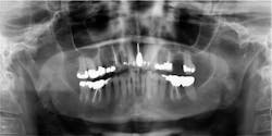
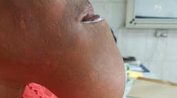
Patient: 71-year-old female
- 5 cm diameter round, greyish-pink mass
- Tissue friable and bleeds easily
- Not tender to palpation
- Located in the right posterior mandible directly behind tooth no. 31
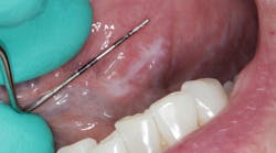
Patient: 77-year-old male
- 6 mm x 8 mm white leukoplakic lesion
- Lesion slightly corrugated with irregular borders
- Not painful to palpation or touch
- Located on left lateral border of the tongue
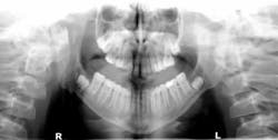
Patient: 14-year-old male
- Patient referred to oral and maxillofacial surgeon to evaluate lower third molars
- Significant, well-circumscribed, expansile radiolucencies around impacted teeth
- Medical history of mental delay
- Recent excision of a basal cell carcinoma from the patient’s chest
Editor’s note: This article first appeared in Clinical Insights newsletter, a publication of the Endeavor Business Media Dental Group. Read more articles and subscribe.
About the Author
Vicki Cheeseman
Associate Editor
Vicki Cheeseman is an associate editor in Endeavor Business Media’s Dental Group. She edits for Dental Economics, RDH, DentistryIQ, and Perio-Implant Advisory. She has a BS in mathematics and a minor in computer science. Early on she traded numbers for words and has been happy ever since. Vicki began her career with Dental Economics in 1987 and has been fascinated with how much media production has changed through the years, yet editorial integrity remains the goal. In her spare time, you’ll find her curled up with a book—editor by day, reader always.


