Pathology case: Differentials that may cross over
Editor's note: Originally published November 29, 2016. Updated June 15, 2024.
Case presentation
A healthy 41-year-old female presented to the office for a routine exam and checkup. Her health history was normal, and she reported no major concerns or issues.
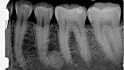
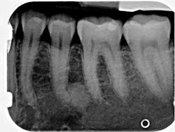
Radiographic examination revealed a large, radiopaque mass just distal to the apical third of tooth no. 20. The area was not tender to palpation, and the tooth tested vital and WNL.
You may also be interested in ... Pathology case: Two related differentials
Differentials
- Periapical idiopathic osteosclerosis
- Mature periapical cemento-osseous dysplasia (PCOD)
- Mature focal cemento-osseous dysplasia (FCOD)
- Hypercementosis
- Condensing osteitis
- Mature fibro-osseous lesions of periodontal ligament origin
The above differentials all share many characteristics, and many cross over. Here is a brief summary of each.
Periapical idiopathic osteosclerosis
- Second most frequently seen periapical radiopacity (after condensing osteitis)
- Idiopathic—emphasizes that the cause of the lesion is unknown
- Located in the periapex of the mandibular first premolar and canine
- Primarily found on healthy, vital teeth
- Asymptomatic, no expansion or palpable lesion, normal mucosa
- Round to irregular in shape; size: a few millimeters to centimeters in diameter
- Varying degrees of density
- Does not have a radiolucent boarder
Mature periapical cemento-osseous dysplasia (PCOD) and mature focal cemento-osseous dysplasia (FCOD)
- Lesions undergo maturation from a radiolucent to a radiopaque stage
- May occur at the apex of vital, healthy teeth
- Completely asymptomatic unless expansion of cortical plates occurs
- Radiographic appearance typically shows a round or oval radiopacity with smooth borders
- Size: 0.5 cm to 2 cm
- Uniformly dense, devoid of a trabecular pattern, and may have a thin radiolucent border
- Root resorption is not characteristic; adjacent teeth may show hypercementosis
Hypercementosis
- Excess formation of cementum on the surface of the root of the tooth
- Altered, club-shaped appearance of the affected root
- Root separates from the adjacent bone by the periodontal ligament
Condensing osteitis
- Occurs in the periapex of a nonvital tooth
- Does not have a radiolucent rim
Mature fibro-osseous lesions of periodontal ligament origin
- Radiolucent rim
- Occurs from trauma; check occlusion!
Diagnosis
Periapical idiopathic osteosclerosis
The lack of radiolucent border and the unknown etiology of the lesion strongly indicate that this lesion is periapical idiopathic osteosclerosis. Since there are no previous radiographs to reference, this diagnosis is not concrete. This diagnosis presents no clinical significance; hence, the patient will be reexamined periodically. Any changes and subsequent treatment will be rendered.
Source
- Wood NK, Goaz PW. Differential Diagnosis of Oral and Maxillofacial Lesions. 5th ed. Mosby Publishing; 1997:460-464.
About the Author
Stacey L. Gividen, DDS
Stacey L. Gividen, DDS, a graduate of Marquette University School of Dentistry, is in private practice in Montana. She is a guest lecturer at the University of Montana in the Anatomy and Physiology Department. Dr. Gividen has contributed to DentistryIQ, Perio-Implant Advisory, and Dental Economics. You may contact her at [email protected].
