Never underestimate the capacity of an infection to stray from the course
Editor's note: Originally published December 6, 2017. Updated December 20, 2024.
Presentation and chief complaint
A healthy 40-year-old male presents to the office with this chief complaint:
“I have some swelling on the lower left side toward the back of my mouth on my jawbone. It’s been there for two days and seems to be getting worse."
Clinical exam
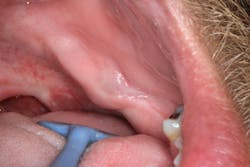
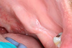
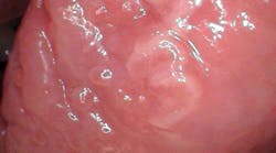
Clinical assessment reveals a fluctuant mass that is tender to palpation on the lingual side of the left mandibular jaw, inferior and lingual to where no. 17 would be, along the palatoglossal arch muscle. The size of the mass is approximately 12 mm x 24 mm, the tissue in the surrounding area is normal in color, and there is no opposing dentition or history of trauma to the area (figures 1 and 2).
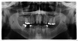
Upon initial assessment, the panoramic radiograph is insignificant (figure 3).
A second opinion and a biopsy
The patient, after having a recurrence of infection and pain, elected to seek a second opinion from another local oral surgeon. This visit resulted in an incision and drainage with a cocktail of amoxicillin/Augmentin/Flagyl antibiotics. A biopsy was also taken that did not come back positive for osteomyelitis. Since there was no true etiology for the lesion (source of chronic infection, immunocompromised system, etc.), consideration was given regarding how to proceed.
A second biopsy
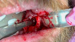
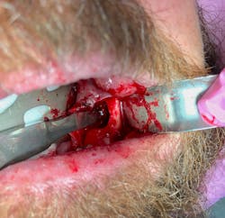
The patient's symptoms improved following the initial drainage; however, another flare-up commenced and it was recommended that a second biopsy be taken. Once the incision was made, a large void in the bone—approximately 2 cm in size—was noted (figure 4).
A large piece of the lower left posterior of the mandible was then resected and sent off for biopsy.
Diagnosis
Acute osteomyelitis
The results came back positive for acute osteomyelitis. Due to the severity of the disease, a PICC line IV was established.
Improvement over the course of the next couple weeks was minimal. The patient—with the support and recommendation of the oral surgeon—elected to go to an academic center for further evaluation, testing, and treatment.
Definition and disease classification
By definition, osteomyelitis is “an inflammatory condition of the bone, beginning in the medullary cavity and Havarian systems and extending to involve the periosteum of the affected area. The infection becomes established in calcified portions of the bone when pus and edema in the medullary cavity and beneath the periosteum compromise or obstruct the local blood supply.”1
Following ischemia, the infected bone becomes necrotic and leads to sequester formation, which is considered a classical sign of osteomyelitis.1 Classification of osteomyelitis is based on the clinical course of the disease:
Acute and secondary osteomyelitis
- Etiology is bacterial; i.e., extraction wounds, foreign bodies, odontogenic disease, periodontal and pulpal infections, infected fractures, etc. The blood supply is oftentimes compromised and can advance rapidly.1,2
- It is characterized by intense pain accompanied by illness, nausea, and overall malaise, which is subsequently relieved with drainage.
- Radiographic presentation is not typically immediately present; however, once the disease progresses and resorbs through the trabecular bone, it will manifest radiographically as a faint, blotchy, mottled lesion with indistinct margins.
- Treatment consists of surgical intervention, drainage, biopsy, and high doses of bacteria-specific antibiotics.2
Primary chronic osteomyelitis
- Rare inflammatory disease that lacks a definitive initiating event; there is a lack of extra- or intraoral fistulas, pus, or sequestration.
- Duration lasts from a few days to several weeks, with alternating episodes of mild to severe pain and swelling.
- Primarily targets the mandible.1
What makes this case challenging is that the patient did not have a clear etiology for the infection and seemed to get progressively worse, despite the initial round of antibiotics from the very first visit.
Take-home points
These are the take-home points to reference from this case:
- Be prepared for the unknown, and don’t underestimate the capacity of an infection to stray from a cut-and-dried course.
- Second opinions are often an insight into additional information that otherwise would not be a consideration. Remember, we are all on the same team.
- Document. Document. Document. While this is important with all medical record keeping, the point is especially driven home when follow-up is needed, referrals are recommended, and multiple providers become involved.
At the time of this publication, the patient was undergoing further testing and receiving definitive care at an academic and health-care facility.
References
- Baltensperger M, Eyrich GK, eds. Osteomyelitis of the Jaws. New York, NY: Springer-Verlag Berlin Heidelberg; 2009:5. doi: 10.1007/978-3-540-28766-7.
- Sapp JP, Eversole LR, Wysocki GP. Contemporary Oral and Maxillofacial Pathology. St. Louis, MO: Mosby; 1997.
About the Author
Stacey L. Gividen, DDS
Stacey L. Gividen, DDS, a graduate of Marquette University School of Dentistry, is in private practice in Montana. She is a guest lecturer at the University of Montana in the Anatomy and Physiology Department. Dr. Gividen has contributed to DentistryIQ, Perio-Implant Advisory, and Dental Economics. You may contact her at [email protected].
