Sharpen your diagnostic skills with this roundup of four pathology cases from DentistryIQ. Your colleagues have shared these cases that you can study and save as a reference as you diagnose future pathology in your clinical exams.
One of the best sources of learning is peer-to-peer. Do you have a case you’d like to share with us? Please email the details to Bethany Montoya, editorial director of Clinical Insights newsletter, at [email protected]. We look forward to featuring you!
Stay tuned for more pathology content from DentistryIQ.
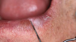
Patient: 60-year-old female
- 5 mm x 24 mm white, corrugated lesion lingual to the acrylic of a fixed hybrid prosthesis
- Lesion not able to be scraped off or removed
- Lesion not painful or symptomatic
- No swellings noted in the sublingual or submandibular lymph node areas
Patient: 40-year-old male
- 12 mm x 24 mm fluctuant mass on the lingual side of the left mandibular jaw, inferior and lingual to where no. 17 would be, along the palatoglossal arch muscle
- Tender to palpation
- Tissue in the surrounding area is normal in color
- No opposing dentition or history of trauma to the area
Never underestimate the capacity of an infection to stray from the course
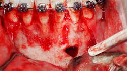
Patient: 77-year-old female
- Two corrugated, irregularly shaped radiopaque masses on the right posterior angle of the mandible
- Each lesion measures approximately 1 inch in length
- Area not tender to palpation
- Health history includes cholesterol and blood thinner medications, type 2 diabetes, history of COPD, high blood pressure, and sensitivity to penicillin
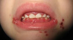
Patient: 16-year-old male
- Multiple lesions salt-and-peppered in a generalized fashion throughout the entire oral cavity
- Lesions are white with red borders
- Gum tissues swollen
- Light palpation/touching of the lesions results in bleeding and pain
Editor’s note: This article first appeared in Clinical Insights newsletter, a publication of the Endeavor Business Media Dental Group. Read more articles and subscribe.
About the Author
Vicki Cheeseman
Associate Editor
Vicki Cheeseman is an associate editor in Endeavor Business Media’s Dental Group. She edits for Dental Economics, RDH, DentistryIQ, and Perio-Implant Advisory. She has a BS in mathematics and a minor in computer science. Early on she traded numbers for words and has been happy ever since. Vicki began her career with Dental Economics in 1987 and has been fascinated with how much media production has changed through the years, yet editorial integrity remains the goal. In her spare time, you’ll find her curled up with a book—editor by day, reader always.


