Pathology with different appearance at different stages
Editor's note: Originally published April 2016. Updated July 2023.
Case presentation
An otherwise-healthy six-year-old male presented to his general dentist. His parents noted that tooth no. 9 had erupted normally following exfoliation of tooth F, but tooth E was still present. They were concerned that the permanent teeth in the area of nos. 7 and 8 might not be developing.
Clinical exam
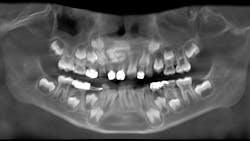
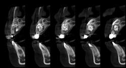
A panoramic film (figure 1) taken by the general dentist revealed a large radiodense area in the region apical to teeth D and E. The patient was referred to an oral and maxillofacial surgeon who obtained a CBCT scan (figure 2).
Differential diagnosis
Clinical and radiographic assessment revealed retained primary teeth D and E, and buccal cortical expansion over the apices of these teeth. A cone beam CT scan revealed a well circumscribed, mixed radiodensity overlying the apices of the primary teeth, adjacent permanent teeth nos. 7 and 8 displaced in a caudal direction, and cortical expansion.
Although the appearance of this lesion should make it readily identifiable, at different stages in its development it may assume a different appearance. The varying appearance at different stages and in varying locations may include these differentials:
- Odontoma
- Ameloblastic fibro-odontoma
- Ossifying fibroma
- Calcifying epithelial odontogenic tumor
- Adenomatoid odontogenic tumor
Treatment
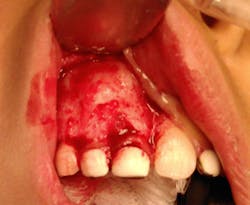
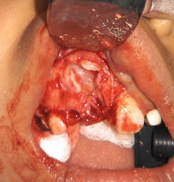
The patient, under intravenous anesthetic, underwent enucleation of the lesion with simultaneous extraction of primary teeth D and E. An intraoral approach was used, the labial cortex was removed, and the mass was removed intact via simple enucleation (figures 3 and 4). The crowns of permanent teeth nos. 7 and 8 were visualized within the cavity of the enucleated mass. No grafting was performed, and the wound was closed primarily in simple fashion.
Final diagnosis
Compound odontoma
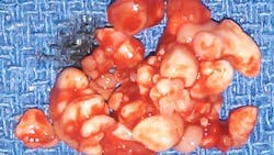
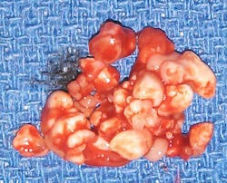
Gross examination of the specimen revealed multiple calcified structures resembling deformed teeth of various sizes within the mass, confirming the clinical suspicion of compound odontoma (figure 5). Two subtypes of odontoma are recognized: compound odontoma and complex odontoma. The compound odontoma is made up of multiple structures of dentin and enamel resembling supernumerary teeth. A complex odontoma consists of a mass of dentin and enamel without the anatomical appearance resembling teeth. The histopathologic report also confirmed the clinical suspicion of compound odontoma.
Prognosis
Each of these lesions is treated via simple enucleation, and there is no incidence of recurrence. In the case noted here, one would anticipate that the disturbance in the eruption pattern of adjacent permanent teeth will require future intervention. Orthodontic consultation and follow-up should be used to appropriately time future intervention and possible exposure of teeth to facilitate forced eruption.
You may also be interested in ... Two patients with the same diagnosis but different treatment
About the Author

R.F. John Holtzen, DMD
R.F. John Holtzen, DMD, is a board-certified oral and maxillofacial surgeon with private practices in Missoula, Montana, and Las Vegas, Nevada. He is a graduate of Temple University School of Dentistry, and completed internship and residency programs at Johns Hopkins University, St. John’s Mercy Medical Center, and MCP-Hahnemann University Medical Centers. He is a previous associate clinical professor at the University of Nevada School of Medicine and has lectured on a variety of topics related to his specialty. Dr. Holtzen is a diplomate of the American Board of Oral and Maxillofacial Surgeons, a diplomate of the American Board of Dental Anesthesiology, and a fellow of the International Association of Oral and Maxillofacial Surgeons.
