Editor's note: Originally published March 2016. Updated June 2023.
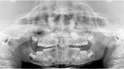
A healthy 7-year-old male presents for his new-patient exam. A radiolucency was noted in the no. 7 area on the panoramic radiograph; subsequent occlusals were referred to for a better view where the lesion appeared to focalize around the coronal portion of no. 7. Furthermore, there was an ill-defined radiopacity within the osseous tissue, under the area where primary tooth D would be located. The area was not tender to palpation nor was there any expansion noted in the vestibular area. Mom reported that the patient had trauma to the area a few years back while playing.
Here are my differentials, definitive diagnosis, and recommended treatment ...
Differential diagnoses
- Dentigerous cyst
- Incisive canal cyst
Definitive diagnosis: benign dentigerous (follicular) cyst
The most common form of pericoronal radiolucencies are dentigerous cysts (DCs).1 DCs are “caused by fluid accumulation between the enamel surface and the reduced enamel epithelium, resulting in a cyst in which the crown is located within the lumen and root(s) outside;”2 the underlying reasoning is unknown. The most common teeth affected are mandibular third molars, maxillary canines, mandibular premolars, and the maxillary canines.1,2 It is not uncommon for a DC to displace the unerupted tooth and often any adjacent teeth. Typically, DCs are painless and slow growing; however, pain will manifest if swelling and inflammation are present.
Recommended treatment
Recommended treatment is complete surgical enucleation. Recurrence is uncommon, but the area must be monitored in the future as there is the potential for residual cysts, ameloblastomas, mucoepidermoid carcinoma, and squamous cell carcinomas to arise from any potentially remaining cells.1,2
In this particular case, the patient was referred to an oral surgeon, who removed a root fragment of what was assumed to be tooth D as well as another piece of random deformed tooth structure. The cyst, along with what appeared to be tooth no. 7 (deformed), was surgically enucleated and the specimen was sent off for biopsy. Results came back as a benign dentigerous cyst. Tooth no. 8—although rotated—appeared to be unaffected.
The patient is being seen by an orthodontist for early interventional orthodontics; treatment options for replacement of tooth no. 7 are being considered as part of his long-term treatment plan.
More pathology cases ...
An asymptomatic, fluctuant mass with a bluish hue
If it's leukoplakic, follow up


