Must-know classifications of dental caries for the national dental hygiene boards
Editor's note: Originally published in 2016. Reviewed for clinical accuracy and formatting in 2022.
Detecting and recording carious lesions is an essential component of the assessment phase in the dental hygiene process of care. Because of its importance, the National Dental Hygiene Board examinations require students to be proficient in detecting and classifying dental caries. So be prepared to be challenged on this topic!
The concepts related to caries may appear in session one (3.5 hours, 200 questions), and the detection of caries using radiographic or photographic images may appear in session two (4 hours, 150 questions with case studies). This article presents two types of caries classifications (G.V. Black classifications, and classifications according to the severity of the lesion) that are most commonly used. Take this chance to fully understand those concepts and train your eyes to detect caries based on radiographic/photographic images. This knowledge will increase your chances at scoring higher on the board exams. If you can recite those classifications as if you are singing your favorite song, you know you are on the right track!
1. G.V. Black Caries Classification (class I to VI)
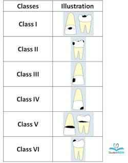
Over 100 years ago, Dr. G.V. Black (1836-1915) developed a system to categorize carious lesions based on the type of tooth affected (anterior or posterior tooth) and the location of the lesion (e.g. lingual, buccal, occlusal, etc.). The six classes of carious lesions according to G.V. Black are as follows:1
- Class I: Cavity in pits or fissures on the occlusal surfaces of molars and premolars; facial and lingual surfaces of molars; lingual surfaces of maxillary incisors (Class I corresponds to surfaces of a posterior tooth you can clinically see—occlusal/lingual/buccal surfaces. Therefore, the interproximal surfaces are not classified as Class I)
- Class II: Cavity on proximal surfaces of premolars and molars (Class II corresponds to surfaces of a posterior tooth you cannot see clinically)
- Class III: Cavity on proximal surfaces of incisors and canines that do not involve the incisal angle (Class III corresponds to surfaces of an anterior tooth you cannot see clinically)
- Class IV: Cavity on proximal surfaces of incisors or canines that involve the incisal angle (Class IV lesion is the larger version of Class III that covers the incisal angle)
- Class V: Cavity on the cervical third of the facial or lingual surfaces of any tooth (Think of the neck of the tooth)
- Class VI: Cavity on incisal edges of anterior teeth and cusp tips of posterior teeth (Class VI corresponds to the very top surface of a tooth)
Review the following example:
Question: According to G.V. Black classification of carious lesions, sealants are placed on permanent molars as soon as they erupt to prevent:
- Class I caries
- Class II caries
- Class III caries
- Class IV caries
- Class V caries
Enamel sealants are generally applied on deep pits and fissures of the occlusal surfaces of posterior teeth. Those areas correspond to the area of Class I carious lesions according to G.V. Black classification (the correct answer choice is 1).
2. Caries classification according to severity
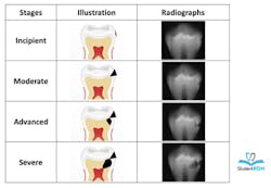
The appearance of interproximal caries can be classified as incipient, moderate, advanced, or severe, depending on the amount of enamel and dentin involved in the caries process.2
Tips for memorization: Imagine a line halfway through the thickness of enamel, and a line halfway through the thickness of dentin. Those lines are the “STOP” points that determine the severity of the carious lesions.
- Incipient: Lesion that extends less than halfway through the enamel
- Moderate: Lesion that extends more than halfway through enamel but does not involve the dentino-enamel junction (DEJ)
- Advanced: Lesion that extends to or through the DEJ but does not extend more than half the distance to the pulp
- Severe: Lesion that extends through enamel, through dentin, and more than half the distance to the pulp
Review the following example:
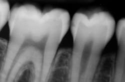
Question: According to the following image, the carious lesion on the distal of the premolar can be classified as:
- Incipient
- Moderate
- Advanced
- Severe
On the radiograph, the dark area protrudes more than half way through the enamel, but does not touch the DEJ. Therefore, this lesion is a moderate lesion. The lesion on the mesial surface of the molar on the other hand can be classified as incipient.
Note: You will be asked to be competent in identifying carious lesions from the radiographs on the case studies in session two. The quality of images may not be very good, but a “magnifier” is available for you to use.
Now we have reviewed some major caries classifications, take this opportunity to master them. These are very important basic concepts that are likely to be emphasized in the National Dental Hygiene Boards. Use this information to successfully pass the exams, and also use them to become an exceptional clinician.
References
- Rashid EG. Operative Dentistry. In: Scheid RC. Woelfel’s Dental Anatomy. 7th ed. Philadelphia, PA: Lippincott Williams & Wilkins; 2007: 432-465
- Interpretation of Dental Caries. In: Iannuci JM, Howerton LJ. St. Louis. MO: Elsevier Saunders; 2012; 402-411


