By Christine Hart
Often when dentists return from a seminar, they want to share new insights into the latest and greatest product that they observed. Sometimes the doctor radiates such enthusiasm that you can clearly anticipate a major change. This was the case at my office a few years ago. Dr. Lewis had been checking out digital radiography systems for a while. Now, he was counting on us to complete his investigation and help make a decision.
Although it is advantageous for our patients and for ourselves to keep up with the latest technology and products, tackling the challenges of “change,” and even research, can seem intimidating. However, if you break it down into smaller parts as we did, you can effectively gather pertinent information that will help make a decision. Then, backed by facts and figures, you can present accurate findings and an action plan to the doctor that can improve and modernize the office.
The only knowledge of digital X-ray we had prior to this point was the occasional dental magazine advertisement that found its way to the office. When the doctor first approached us with the idea, we had many questions. After all, we take X-rays throughout the day, every day. We wanted assurance that:
- digital radiography was going to be cost-effective
- we would be able to learn how to use the digital software quickly
- our patients would embrace and appreciate the technology
- we would choose the best system for our office.
We started to gather information. First, we looked at economics. We knew that there was not only the cost of film and chemicals, there were mounts and envelopes, duplication film, and processor cleaning supplies. There was also the disposal of hazardous chemicals regulated by our state, and the time involved in developing film and cleaning the processor.
From our practice management software, we were able to find out how many pieces of film and, from the number of series, the total of mounts we used in a given period. We determined how much we were spent on duplicating film, chemicals, and processor cleaners using our supplies invoices. We calculated the cost of these items for the average month, and then added one-third of our quarterly chemical disposal fee.
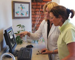
FIGURE 1. Large image allows patients to see and understand
After obtaining this monthly total, we compared it to the proposed monthly expense that we would have with a digital X-ray system, including some computer modifications to our treatment room workstations. Numbers don’t lie.
We were shocked to find out how much we could save by going digital. I challenge you to do your own computations - you’ll probably be surprised, too!
The next topic, the learning curve of computerized X-ray software, was addressed by a field trip to a local periodontal office with a digital system to see and understand the step-by-step procedure of capturing and using digital images. The office staff was kind enough to explain how they initially implemented the process into their daily routines, and to show us some of their favorite clinical and diagnostic features. Observing this ‘real-life’ clinical situation greatly influenced our decision to go forward.
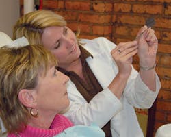
Our continued research showed some real advantages for our patients, such as lower radiation levels and greater understanding of diagnoses. It’s much easier to use large images on a monitor (Fig. 1) rather than small pieces of film (Fig. 2) to explain conditions and procedures to patients. Based on these findings, and because patient involvement is very important in our practice, we knew these benefits would add up to a great improvement.
Armed with our new knowledge, we all decided that we definitely should make the change to digital. Now, we only needed to narrow the field on which system would best meet our needs. Ultimately we chose one that has the ADA seal stating that it’s equivalent to film, as we did not want to compromise diagnostics.
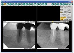
FIGURE 3. Enhanced images for better diagnostic capability
Not only are the images excellent quality, but because of some of the diagnostic tools we are now able to see things we could not see on film. For example, there are enhancement features that allow us to sharpen and invert the lights and darks on an image (Fig. 3). This tool has allowed us to verify caries and find cracks along the root of the tooth.
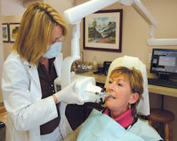
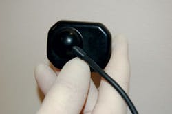
Instead of using multiple-sized sensors, our chosen system has a single-sensor solution that because of its design, allows us to take images quickly in any area in all sizes of mouths (Fig. 4). It is also very comfortable for the patient (Fig. 5) and the software is very “user-friendly,” even for novices. If you know how to take an X-ray and click a mouse, you can use this system.
Our digital X-ray instructor was a practicing hygienist who became certified to teach for the company. She also used the product in an office - she knew what it was like to make the transition from film to digital. We were able to take images with her by our side. The software was so easy to learn that we went “live” with our system the same day, never using film again! Not only did we stop purchasing film, chemicals, and supplies, we stopped wasting the time related to film processing.
Eliminating environmental waste is another advantage that is very important to us. None of us likes working with X-ray chemicals, including the specialized cleaners for the processor. The MSDSs let us know how caustic these chemicals are, definitely not good for the office environment. I personally am glad that we are not contributing to the toxic waste in our global environment. Besides this benefit, we have eliminated the cost associated with disposal.
FIGURE 5. Beveled corners and rounded edges allow for comfort
An initial concern we had was sharing one sensor among five clinicians working at the same time. We found this to be no problem at all! The digital images are on the screen so quickly that we soon realized we used to wait much longer for the darkroom to become available than we ever wait now for the sensor. We also store our sensor and holders in a container in a central place so that everyone can locate them quickly.
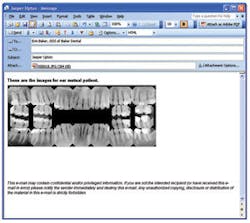
Our workflow and efficiency have improved as a result of our acquiring digital X-ray. We no longer have the need to duplicate images for other offices or insurance companies. It is so simple; all we do is print the image on paper and reprint it if they need another copy, or better yet, we can e-mail it to them (Fig. 6). Also, because the images are dated and have the tooth number on them, it makes it very easy for the front office staff to locate a requested image.
FIGURE 6. E-mail images for quick communication with colleagues
Several years after we implemented digital X-ray, we made another big change for the better. Utilizing our system, along with our practice management software, has allowed us to become paperless. The time we save pulling and filing charts is a great time-saving benefit.
Lastly, as we expected, our patients appreciate 60 percent reduction in radiation. Now, we enlarge an image on the screen not only for our viewing, but also to help our patients understand sufficiently to become more involved with their treatment. They compliment us on being “cutting edge.” We feel we are providing the best possible care to our patients.
When we built our new office, our digital X-ray system was an important aspect of the new surroundings - we wouldn’t have moved without it! Plus, having this technology allowed us to use
the square footage that would normally be wasted on a darkroom, to be used for a treatment area - a much better use of space!
I feel fortunate to be part of an office that values the opinions of all its team members when making changes to the practice. Through education, we faced our fear of change and were able to take the positive step to go forward with our digital X-ray system. We now have a wise investment, one that is good for our patients, our team, our practice, and our environment. Are you ready for a change for the better?
Biographical Sketch
Christine Hart has 19 years clinical and administrative experience in the field of general dentistry, as well as a strong history in computer learning. She is currently a 12-year team member with Dr. Michael R. Lewis in Rochester, NY. Christine is happy to answer your questions. She can be reached at [email protected].

