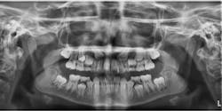
Impacted canines are a common orthodontic problem that is treated more easily when recognized early. Making the correct diagnosis is essential. Use the following criteria to help determine if there is a problem with canine eruption.
Assess the patient's dental age. Typically canines erupt around age 12. It is not unusual for boys to be delayed by one year and occasionally by two years. A girl who has canine eruption delayed by more than one year may have an impaction problem.
Look at the stage of development of the opposite canine. If one canine is fully erupted and the other is nowhere in sight, there may be an eruption problem.
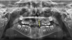
Evaluate the positions of the unerupted canines on a panoramic radiograph for all patients at ages eight and nine. Whenever the crown of the canine crosses the long axis of the lateral incisor, as shown in Figure 1, the canine may become impacted.
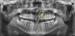
Look at the size of the follicle around the unerupted canine. Canines that are having problems with eruption tend to have larger than normal follicles, as seen in Figure 2.
Take a panoramic radiograph on all patients with missing maxillary lateral incisors and pegged lateral incisors on a regular basis and evaluate the maxillary canine positions. There is a genetic component to missing and pegged maxillary lateral incisors. There is a high correlation between missing and pegged maxillary lateral incisors and impacted canines.
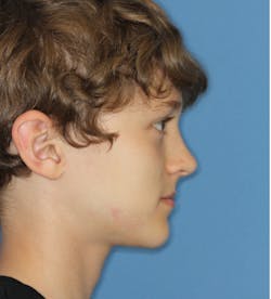
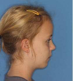
Monitor all class III patients for impacted maxillary canines. There is a high correlation between a class III skeletal pattern and impacted maxillary canines, particularly in patients who are maxillary deficient. Patients who are maxillary deficient tend to have a class I molar relationship at an early age (six years old) and minimal overjet. When viewing the profile, it appears straight, as seen in Figure 3. They may also have anterior and posterior crossbites. Normally, six-year-olds should have an end-to-end class II molar relationship with 3–4 mm of overjet, and they should have a class II, mandibular-deficient profile, as seen in Figure 4.
Patients with ectopically erupting maxillary canines are at risk for root resorption on the adjacent lateral incisors. It is a misconception that lateral incisor root resorption is the result of orthodontic treatment. Lateral incisor root resorption resulting from orthodontic forces is controllable and is usually minimal. Patients presenting with root resorption at the start of orthodontic treatment will experience the most root resorption. This is due to the poor position of the canine and resorption of the lateral incisor root by the canine, not by orthodontic forces. It is unusual to see root resorption as severe as that of the nine-year-old patient show in Figure 5; however, such severe root resorption may be avoided by recognizing a potential problem early.
Editor's note: This article first appeared in the Product Navigator. Click here to subscribe. Click here to learn how to submit an article.
