Like it or not, lasers in dentistry are here to stay. Many clinicians have embraced lasers for both hard- and soft-tissue applications. The soft-tissue lasers used as adjuncts for nonsurgical periodontal scaling and root planing also have surgical periodontal applications. These surgical applications have multiple names and acronyms: laser periodontal therapy, or LPT, is a common reference to the surgical approach with lasers, and the trademarked name Laser-assisted New Attachment Procedure, or LANAP, is also commonly used in reference to laser periodontal surgery. These surgical procedures do not require a traditional flap. It is assumed that patients are more willing to complete periodontal treatment when it is completed with a minimally invasive approach, such as laser periodontal surgery.
Within the dental community, there are debates concerning the efficacy of laser periodontal therapy. Many studies can be found both in both support and rejection of these procedures. However, there are limited firsthand clinical reports and cases identifying the protocols that have had the greatest impact on laser surgical success.
The differences between a nonsurgical and surgical approach can include multiple factors beyond the laser settings. The following steps have been noted in the literature and recommended by clinicians: evaluation of occlusion and adjustment as indicated, closing open contacts, and fabrication of an occlusal guard. Some of these steps, if not all, have become part of many LPT protocols. Although each one of these factors contributes to surgical success, none of them directly address the primary etiology of infectious periodontal disease.
According to the American Dental Association Council on Scientific Affairs, therapies intended to arrest and control periodontitis depend primarily on effective root debridement. Disruption of biofilm and removal of biofilm-harboring calculus is the gold standard of periodontal care, both with a conventional flap and nonsurgical scaling and root planing. Effective debridement should carry as much weight in minimally invasive, laser-assisted periodontal surgery as it does in other treatment modalities.
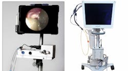
In 2014, Osborn et. al found that the use of an endoscope resulted in significantly less residual calculus on the root surface, compared to tactile evaluation during scaling and root planing.1 To date, no studies have compared the immediate and long-term results of LANAP performed with and without a dental endoscope.In our periodontal office, the doctor has been performing LANAP surgery for 10 years. This protocol includes closing open contacts, adjusting occlusion, and recommending use of a nightguard. For the past three years, LANAP patients have had the option of adding use of the dental endoscope to their procedures. A dental endoscope allows for magnified, subgingival visualization through the use of a fiber optic. This allows the clinician to inspect and debride the root structure, rather than relying on tactile inspection.
For this article, a small sample of cases were reviewed and compared to see if the addition of the endoscope made a difference in initial and long-term results. This review is not intended to be definitive proof that an endoscope should be utilized with laser periodontal surgery. The intention of this article is to begin a discussion about the role of dental endoscopes in minimally invasive, laser periodontal surgery.
Ten patients' cases were reviewed, five of which involved LANAP only and five of which involved LANAP plus debridement with a dental endoscope. Following the current American Academy of Periodontology guidelines, all of these cases would be classified as generalized stage III, grade B. All of the patients had a history of smoking, and eight were currently smokers.
Each patient was evaluated surgical results, no earlier than nine months post-op. These cases were beyond two years postsurgery and had been following a three-month recall schedule since treatment. Table 1 compares the two groups.
Table 1: A comparison of LANAP surgical results with and without an endoscope
| Mean pocket reduction at 9 months post-op | Number of bleeding sites at 9 months post-op | Number of pocket depths greater than 3 mm at 24 months post-op | Number of bleeding sites at 24 months post-op | |
| LANAP only | 4.8 mm | 2 | 12 | 18 |
| LANAP and endoscope | 6.2 mm | 0 | 4 | 0 |
Even after treatment, patients' periodontal health is fragile. Our ultimate goal as practitioners is to reduce or eliminate the signs of inflammation and halt the progression of periodontal breakdown. These findings suggest that using a dental endoscope in conjunction with LANAP results in not only a greater reduction of pocket depths than LANAP alone, but also shows continued elimination of the key indicator of inflammation, bleeding upon probing.
This review is in no way a scientific representation of the benefits of endoscope use with a laser surgical protocol, and there are many reasons why the results vary between the two approaches. This is, however, a call to practitioners, educators, and researchers to further examine the likely benefits of using a dental endoscope with laser periodontal surgery. If we are recommending occlusal guards, closing open contacts, and adjusting areas of traumatic occlusion, perhaps we should also be ensuring a more complete decontamination and debridement of the root surfaces. After all, that is the key to success.
Reference
1. Osborn JB, Lenton PA, Lunos SA, Blue CM. Endoscopic vs. tactile evaluation of subgingival calculus. J Dent Hyg. 2014;88(4):229-236.
Nicole Fortune, MBA, RDH, is a registered dental hygienist who works full-time in a periodontal office. She is a recognized expert in training dental professionals in areas of periodontics, including peri-implantitis and dental endoscopy. Nicole has delivered educational presentations to numerous institutions, clubs, and professional groups across the country. Nicole earned her hygiene degree and her bachelor's degree from the University of Vermont and her master's degree from Champlain College.


