Editor's note: Originally published March 31, 2015. Updated February 2023.
Case presentation
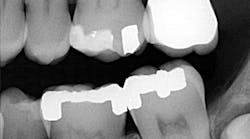
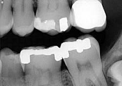
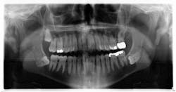
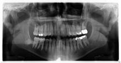
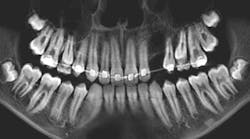
A healthy, 38-year-old male presented to the office for a comprehensive exam. His blood pressure was within normal limits, and no medications were being taken. Bitewing radiographs were taken, and it was noted that there was a cystlike lesion on what appeared to be a mesial impaction of no. 17. A panoramic radiograph indeed revealed a large, radiolucent/cystlike lesion surrounding a mesial/inferior-positioned no. 17. The widest point of the cyst measured approximately 3 cm. The patient reported knowing about the lesion but was not inclined to have it removed because it was not bothering him. The area was not tender to palpation; tooth no. 18 had normal pocketing and tested normal to cold, percussion, and bite.
You might also be interested in…
Periodontal abscess or gingival abscess? A clinical case report
Oral pathology case: The common but not-so-common radiopacity
The “non-bothersome” soft-tissue lesion
Diagnosis and treatment
Dentigerous cyst. A dentigerous cyst is “an odontogenic cyst that surrounds the crown of an impacted tooth, caused by fluid accumulation between the reduced enamel epithelium and enamel surface, resulting in a cyst in which the crown is located within the lumen and root(s) outside.”1 Typically, dentigerous cysts remain asymptomatic, but large growths have been known to produce swelling and pain. The most common teeth involved are the unerupted maxillary and mandibular third molars or maxillary cuspids.1 The highest incidence occurs during the second and third decades of life.2 The recommended treatment is complete enucleation, which reduces the possibility of potentially dangerous cells remaining in the region after surgery to form residual cysts, ameloblastomas, or other lesions.2
In this particular case, the patient was referred to an oral surgeon and the lesion was removed. Histology confirmed the diagnosis, and the patient has been seen on a regular basis without any complications or recurrent lesions. The bone has filled in nicely and no. 18 is vital. The coronal portion of no. 32 was removed due to the wrapping of the roots around the inferior alveolar nerve.
References
1. Sapp JP, Eversole LR, Wysocki GP. Contemporary Oral and Maxillofacial Pathology. Mosby Publishing; 1997;42-43. 2. Wood NK, Goaz PW. Differential Diagnosis of Oral and Maxillofacial Lesions. 5th ed. Mosby Publishing; 1997;283-285.
