Case presentation and history
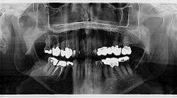
A 78-year-old male was referred to the oral and maxillofacial surgeon to evaluate a radiodense area discovered as an incidental finding on a panoramic radiograph (figure 1). The patient is a healthy, active individual with a medical history remarkable only for well-controlled hypertension and mild gastroesophageal reflux. His medications include lisinopril and omeprazole, and he has no known drug allergies.
The area noted on the panoramic film appeared as multiple well-defined radiodense objects inferior to the condyle and anterior to the right condylar neck.
The patient reported a history over many years of a popping noise emanating from the right temporomandibular joint (TMJ) on function and occasional discomfort in the same area. The popping eventually subsided and was replaced with crepitance and less frequent, but still occasional, discomfort. The patient denied any limitation of movement as well as a history of open or closed lock. He did not recall any history of facial trauma.
Comprehensive exam and assessment
Clinical examination revealed a healthy adult dentition with a stable class 1 occlusion. The mandible had a normal range of motion with maximal opening of 44 mm, protrusion of 8 mm, right and left lateral excursive movements of 8 mm, and no deviation on opening. Subtle crepitance was palpable over the right TMJ, but there was no tenderness of either joints or facial musculature. The differential diagnosis at this point included osteoarthritis with benign osteophytes, synovial chondromatosis, and calcium pyrophosphate deposition (CPPD) disease.
More pathology cases:
Synovial chondromatosis of the TMJ is also a rare, benign, proliferative disorder that results in the production of cartilaginous nodules within the joint space of synovial joints. Over time the nodules detach and calcify, moving freely within the joint. Patients with synovial chondromatosis often present with pain, trismus, and swelling of the joint. Radiographic appearance typically displays a heterogenous mass within the joint space associated with prominent joint effusion.3,4
TMJ osteoarthritis results from a spectrum of factors that produce both inflammatory changes in the joint and the consequences of mechanical overload. The typical patient experiences symptoms ranging from pain, to swelling and limitation of function, to painless joint noise, or, frequently, no symptoms at all. Radiographic examination reveals degeneration and sclerosis of the joint structures such as bony remodeling, degeneration and displacement of the meniscus, and occasionally proliferative bony changes.5 The mechanical overload component may produce bony proliferation, which can ultimately separate and form free-floating osteophytes.6 This condition is essentiallyDiagnosis
Definitive diagnosis in the case cannot be ascertained without histopathological evaluation of the radiopaque masses noted on the films. The presumptive diagnosis for the patient described in this case is benign osteophytes resulting from osteoarthritis of the TMJ. Given the benign appearance at present, coupled with the open joint or arthroscopic procedure required for definitive diagnosis, a decision was made to manage this case with close observation for the foreseeable future.7 In the event the radiographic appearance becomes more remarkable or the patient develops significant symptoms, the option of surgical intervention will be revisited.
References
1. Kwon KJ, Seok H, Le JH, et al. Calcium pyrophosphate dihydrate deposition disease in the temporomandibular joint: diagnosis and treatment. Maxillofac Plast Reconstr Surg. 2018;40(1):19. doi:10.1186/s40902-018-0158-0
2. Canhão H, Fonseca JE, Leandro MJ, et al. Cross-sectional study of 50 patients with calcium pyrophosphate dihydrate crystal arthropathy. Clin Rheumatol. 2001;20(2):119-122. doi:10.1007/s100670170081
3. von Lindern JJ, Theuerkauf I, Niederhagen B, Bergé S, Appel T, Reich RH. Synovial chondromatosis of the temporomandibular joint: clinical, diagnostic, and histomorphologic findings. Oral Surg Oral Med Oral Pathol Oral Radiol Endod. 2002;94(1):31-38. doi:10.1067/moe.2002.123498
4. Balasundaram A, Geist JR, Gordon SC, Klasser GD. Radiographic diagnosis of synovial chondromatosis of the temporomandibular joint: a case report. J Can Dent Assoc. 2009;75(10):711-714.
5. Zarb GA, Carlsson GE. Temporomandibular disorders: osteoarthritis. J Orofac Pain. 1999;13(4):295-306.
6. Sadaksharam J, Khobre P. Osteophytes in temporomandibular joint, a spectrum of appearance in cone-beam computed tomography: report of four cases. J Indian Acad Oral Med Radiol. 2016;28(3):289-291.
7. Moses JJ, Hosaka H. Arthroscopic punch for definitive diagnosis of synovial chondromatosis of the temporomandibular joint. Case report and pathology review. Oral Surg Oral Med Oral Pathol. 1993;75(1):12-17. doi:10.1016/0030-4220(93)90398-n


