Case history and presentation
A 66-year-old male presents for his routine exam and periodontal maintenance visit. His health history includes medications for blood pressure and cholesterol.
Clinical assessment
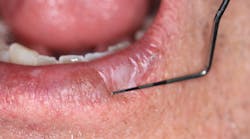
Upon performing a head, neck, and oral cancer screening, a 6 mm x 9 mm lesion was noted on the lower left side of the patient's lip. The lesion was irregular in shape and white in appearance (figures 1 and 2). There was no sluffing or pain. Upon inquiry, the patient stated that the lesion appeared approximately two months ago and did not go away. He did not recall any trauma to the area either.
Figure 1: Leukoplakic lesion on lower left lip
Figure 2: 6 mm x 9 mm irregular-shaped lesion appears white
What is your overall assessment of the lesion and what would you recommend to this patient, keeping in mind that lack of pain is often not enough to nudge a patient into seeking a second opinion or a definitive diagnosis? Furthermore, what are your differentials?
Send your responses to [email protected]. Next month, we will present the final diagnosis and recommended treatment for this oral pathology case.
Read more pathology articles at this link.
CALL FOR PATHOLOGY CASES
Do you have an interesting oral pathology case you would like to share with Breakthrough’s readers? If so, submit a clinical radiograph or high-resolution photograph, a patient history, diagnosis, and treatment rendered to [email protected].
Editor's note: This article originally appeared in Breakthrough Clinical, a clinical specialties newsletter from Dental Economics and DentistryIQ.


