Obstructive salivary gland disorders and treatment options: what dentists need to know
Until recently, patients in the United States who suffer from obstructive salivary gland disorders had few options for relief. But today, advances in science and medical device technology are providing a variety of innovative and effective minimally invasive treatment options for patients.
The major salivary glands of the human body produce approximately one quart of saliva each day. Saliva plays a crucial role in digestion, swallowing and speaking, and in protecting teeth against bacteria and decay. The most common problems in these vital glands occur when the ducts become blocked and saliva cannot drain. In fact, the latest data show that approximately one in every 5,000 people will develop some type of obstructive salivary gland disorder. (1)
The most common of these disorders is sialolithiasis, or salivary stones, a condition that can affect patients of all ages. Sialolithiasis occurs in males twice as often as in females. Recently published literature suggests that the autoimmune disorder Sjogren’s syndrome, which affects more than two million patients in the United States, can cause inflammatory obstruction of salivary flow. And with 40,000 new cases of thyroid cancer being diagnosed each year, radioiodine-induced sialadenitis is just now starting to be recognized by researchers.
ADDITIONAL READING |Chairside Salivary Diagnostics for Oral Diseases
Patients with obstructive salivary gland disorders, including sialolithiasis, often contact their dentist first when they experience discomfort or other oral health issues. Because dental professionals are the first line of defense in identifying these disorders in patients, they must recognize the warning signs and know the treatment options that are available.
Some of the warning signs are pain and swelling in the cheek or under the jawline while eating. Pain and swelling may be intermittent or can suddenly get worse before mealtimes and then slowly get better. Patients may also experience a foul taste in the mouth when the duct tightens or is blocked by a stone. Obstructive salivary gland disorders can progress to infection, high fevers, severe pain, and increased swelling. Many patients are embarrassed to discuss this condition with their physician. But when left untreated, the disorder can cause debilitating quality-of-life issues.
ADDITIONAL READING |Inflammation of the gingiva: The other reasons besides the obvious
Patients who have recurring episodes of sialolithiasis may require open surgery, which is performed by otolaryngologists and oral and maxillofacial surgeons. Open surgery is the most common procedure that is performed on submandibular glands. (2) Parotid salivary gland resection is a less common open surgery because of its higher incidence of postoperative complications, such as facial muscle weakness or loss of voluntary facial muscle movement. (3) Other complications from parotid salivary gland resection can include postoperative pain and potential airway complications from excessive bleeding in the mouth.
Because of the complications from open surgery, many otolaryngologists and oral and maxillofacial surgeons are now adopting salivary endoscopy, or sialendoscopy, as a new treatment modality.
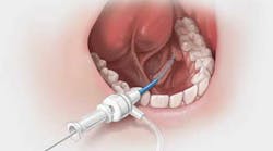
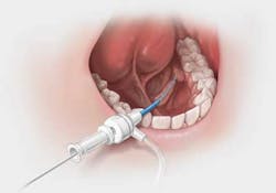
Sialendoscopy is a minimally invasive procedure for visualizing and treating these disorders directly through the salivary ducts. Physicians can perform this procedure in the office through a small endoscope, which is inserted into the salivary ducts to visualize, diagnose, and treat inflammatory and obstructive conditions, such as stones. Stone retrieval baskets and graspers often can be used to remove a stone after successful ductal access is achieved. Complications may include salivary duct wall perforations, basket entrapment, postoperative infections, temporary lingual nerve paresthesia, and ductal stenosis.Click on the photo to view a sialendoscopy procedure animation
Otolaryngologists and oral and maxillofacial surgeons may use these devices during sialendoscopy to access the very small salivary ducts — which is one of the most challenging aspects of the procedure — and to manipulate and remove stones through the endoscope. As patients and physicians continue to seek newer treatments for this painful disorder, sialendoscopy may be considered one option for eliminating open surgical procedures that carry undesirable risks.
References
1. Cashin-Garbutt A. Obstructive salivary gland disease treatment: an interview with Thomas Cherry, Cook Medical. News Medical website. http://www.news-medical.net/news/20130419/Obstructive-salivary-gland-disease-treatment-an-interview-with-Thomas-Cherry-Cook-Medical.aspx


