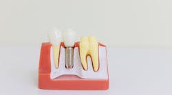
Your client has had some trouble with their implant for some time and you have done your best to instruct them on proper care including brushing, flossing, WaterPik, sulca brushes—all to no avail. Every radiograph is showing continued loss of bone height. The dentist informs the client that the implant “has failed” and will have to be removed. The client then turns to you and asks . . .
“Why did my implant fail?”
“It’s not hurting. Can we try to save it?”
“Will I be able to get another implant?”
“If I get a new implant will it fail as well?”
If your dental team is unsure how to respond to these questions, you aren’t alone.
While most implants succeed, the answer to the question “Why do implants fail?” is complicated. “The scientific literature regarding the risk factors for implant failure is limited . . . the treatment of biologic complications and failing implants lack systematic scientific validation and is based mainly on empirical experience and inference from in vitro findings on a trial and error basis.”1 From the evidence gathered, common risk factors and profiles do emerge: “the [type of] implant device, surgical procedure, anatomy, systemic health, occlusion, microbial biofilm, host immuno-inflammatory response and genetics.”1 However, many of these can be minimized if all the factors that lead to implant failure are considered when planning treatment.1 With the success and popularity of implants steadily growing as a long-term standard treatment option, so grows the need to understand factors combining and contributing to implant failure.
There are two main types of implant failure, early and late. Early failures occur within the first three to four months of implant placement and are due to a lack of osseointegration. When considering placement or replacement of a dental implant, the clinician and client must examine risks for early failure. These include:
- Autoimmune diseases (e.g., diabetes, rheumatoid arthritis) in which the body rejects the implant as a foreign body
- Bisphosphonate medications that may affect the growth of bone support after placement
- Poor blood supply due to the effect of medications or smoking
- Overloading, when bite pressure is focused on the implant instead of surrounding teeth, occurs occasionally when the crown was not seated well, the implant is in a posterior location and the bite force is heavy, the client clenches, or an implant bridge span is too long
- Rejection of a foreign body, when the body will not allow integration and forces the implant out
- Allergies to implant components can occur. Implants are a titanium alloy and contain minor amounts of nickel that may trigger sensitivities. The client may complain of a burning or tingling at the site.
- Various implant types are now available, and all have their pros and cons. Every client is unique in their situation and this must be considered when choosing the implant type. Machined surfaces have performed poorly compared to other options available, including roughened surfaces, titanium oxide coated, chlorhexidine coated, ceramic coated, or completely ceramic. All have their benefits in certain conditions, but all are striving for better osseointegration in a maintained clean field.
- Factors relating to the surgical procedure: Malposition of the implant in the basal layer of bone can lead to loosening or various parts breaking. Especially during direct placement after tooth removal, the site must be cleaned of all necrotic or diseased tissue, the dense cortical bone of the socket walls removed while being mindful of not creating too much heat, the site irrigated and disinfected, and the implant itself must be sterile. One study noted that surgical trauma and bone quality and quantity were the most important etiological factors involved in early implant failures.2
- Infection: Factors such as other areas of gingivitis and periodontitis, especially on teeth close to an implant, as well as the client’s general oral hygiene, must be considered. Foreign objects such as rough cement, or food debris, are also known to be some causes of inflammation leading to implant failure shortly after placement.
Exploring long-term failure, these are problems that occur after osseointegration, and are many of the same reasons people lose natural teeth. Risk factors such as poor oral hygiene, clenching and grinding, smoking, general poor health, excessive load, and the amount or quality of bone, are all issues that can all lead to failure of an implant even after successful integration.
A 2015 systematic review by a working group of periodontal researchers and experts found a prevalence for peri-implant mucositis of 43% and for peri-implantitis of 22%, and
“lack of regular supportive therapy in patients with peri-implant mucositis was associated with increased risk for onset of peri-implantitis . . . patient-administered mechanical plaque control (with manual or powered toothbrushes) has been shown to be an effective preventive measure; professional intervention comprising oral hygiene instructions and mechanical debridement revealed a reduction in clinical signs of inflammation; [and] adjunctive measures (antiseptics, local and systemic antibiotics, air-abrasive devices) were not found to improve the efficacy of professionally administered plaque removal in reducing clinical signs of inflammation.”3
In other words, prevention of disease, and control of peri-implant mucositis, is typically focused on early detection and intervention from the dental hygienist through physical and educational means, and this centers around control of inflammation through plaque removal. The working group suggests that there is little evidence to show the benefit of adjunctive services. However, detailed research does suggest that, generally, if only the smooth surface of the abutment is exposed and the rough anatomy of the implant is not accessible, hand debridement is just as successful as other means. However, when the rough surface meant for osseointegration is exposed, though hand scaling is still required to remove larger pieces of calculus, “the air polishing device did best at reaching all implant areas including the tips of the threads and between threads.”4
It seems that there is still no consensus on which scalers are the best to use. Resin or nylon curettes and plastic-tipped ultrasonics are said to have the least impact on the implant surface compared to steel or titanium scalers. However, plastic instruments also leave behind particles on the rough implant surface, whereas there is less chance of this with the other scaler types. Completing removal of remaining calculus and biofilm is still best achieved, with the least amount of alteration to the implant surface, with an air abrasive device and glycine or erythritol powders.
If peri-implantitis reaches a moderate level of 3 to 4 mm of CAL (after the initial osseointegration level has settled), we must decide to consider a treatment beyond preventive debridement. Implant removal before moderate or severe bone loss occurs is still often a recommended intervention. However, new treatment methods are being investigated and have opened up the possibility of reversing peri-implantitis. Surgical debridement of the pocket and the disinfection of implant body are imperative to stopping the disease process. Interventions should include periodontal surgery to open the flap and reveal the implant and defective bony margin. Next, removal of any necrotic bone or tissue, calculus, and biofilm, to disinfect the implant body and cleanse the site may be done. This can be completed through chemical, laser, or erythritol/glycine air abrasion methods. All have their benefits or drawbacks but chemical and air abrasion methods have the most positive results compared to laser, as issues of heat and proper decontamination of the tissue arise, impeding proper healing. Again, factors that led to peri-implantitis must be considered and resolved for this treatment to be successful.
If an implant must be removed, a replacement is often considered: “Implant replacement is a reasonably feasible option for scenarios of early and late implant failure. However, modifiable risk factors must be controlled before proceeding for implant replacement.“5 This includes the age of the client, their medical status, smoking, oral hygiene, financial status, the desires of the client, and the anatomical viability of the site. “An overall implant survival rate of 86.3% after retreatment suggests that most initial implant failures are likely attributable to modifiable risk factors, such as implant architecture, anatomic site, infection, and occlusal overload.”6
In the end, their seems to be little consensus in the research, and therefore little consensus on determining if an implant will fail with any certainty, and with all the factors involved in it is plain to see why. We are making headway however, and an understanding of risk factors, and combinations of risk factors, can still guide us in helping our clients with their decision-making process when it comes to how to manage a failing implant.
By understanding the risks that can lead to implant loss, dental hygienists can be equipped to intervene during the early stages of peri-implant mucositis and peri-implantitis, educate our clients on the factors that likely led to peri-implantitis, collaborate on whether these factors can be managed well at home and in the dental office, or to consider a periodontist referral. By understanding our client’s unique risk factors, we are equipped to help them sort through the information and decide what option may be best for them—to try to save an ailing implant, consider removal of the implant and replacement, or consider another option for tooth replacement.
References
1. Shieh J. Biomechanical implant failures management of an implant failure: A case report. Oral Health Group website. https://www.oralhealthgroup.com/features/biomechanical-implant-failures-management-of-an-implant-failure-a-case-report/. Published February 5, 2019.
2. Palma-Carrió C, Maestre-Ferrín L, Peñarrocha-Oltra D, Peñarrocha-Diago MA, Peñarrocha-Diago M. Risk factors associated with early failure of dental implants. A literature review. Med Oral Patol Oral Cir Bucal. 2011 Jul 1;16(4):e514-7.
3. Jepsen S, Berglundh T, Genco R, et al. Primary prevention of peri-implantitis: managing peri-implant mucositis. J Clin Periodontol. 2015;42 Suppl 16:S152-7. doi:10.1111/jcpe.12369.
4. Steiger-Ronay V, Merlini A, Wiedemeier DB, Schmidlin PR, Attin T, Sahrmann P. Location of unaccessible implant surface areas during debridement in simulated peri-implantitis therapy. BMC Oral Health. 2017;17(1):137. doi:10.1186/s12903-017-0428-8.
5. Zhou W, Wang F, Monje A, Elnayef B, Huang W, Wu Y. Feasibility of dental implant replacement in failed sites: A systematic review. Int J Oral Maxillofac Implants. 2016;31(3):535-45. doi:10.11607/jomi.4312.
Amy L. Dion, BSc, RDH, is an independent dental hygienist and practice owner of 10 years. Her experiences include working in cosmetic, implant, and oral cancer recovery dental offices, and teaching dental hygiene in the clinical setting. She currently practices within an implant denture clinic and from her home office. Her passion is bringing dental prevention to her clients by means of a holistic experience, in-depth risk assessments, and use of innovative products for clinical and home use. She is a member of the CDHA and ODHA. Contact her at amydionrdh.com.


