Oral rest posture: A key piece of the obstructive sleep apnea puzzle
Dental professionals are missing the chance to address obstructive sleep apnea before it even begins.
In the past few years, the subject of the airway in relation to dentistry has been increasingly discussed. Obstructive sleep apnea (OSA) and snoring are hot topics right now. There seems to be a call to action for all dental professionals to assess our patients for snoring and sleep apnea. Mandibular advancement devices and take-home sleep studies are definitely trending in the dental world.
The spotlight on this issue is necessary and important. But rather than focus all of our attention on the treatment modalities that dental professionals can offer for snoring and sleep apnea, what if we shifted our focus to how we can help to prevent these issues in the first place? What if we started to look at our younger patients through a new lens, and learned how to evaluate even the slightest changes in growth and oral muscle function? What if we had a clear understanding of the impact that these changes could have on the future health and wellbeing of our patients?
There are many factors that influence the development of snoring and sleep apnea. Diet, weight, and neck circumference often play a role in development of OSA. However, according to prominent ENT and sleep apnea expert Steven Park, MD, OSA is actually a craniofacial problem. (1) Patients with smaller jaws have smaller airways. Constricted arches leads to less volume inside of the mouth, which lessens the space necessary for good tongue posture. The maxilla includes the nasal floor and is not only the roof of the mouth. Therefore, a high narrow arch leads to a narrowing of the side to side distance of the nose. (1) A high palatal vault may lead to deviation of the nasal septum. (1)
When making the connection between developing oral and facial structures in children and snoring and OSA in adults, we need to look at a factor that often goes unnoticed: oral rest posture (ORP). ORP refers to the proper position of our muscles when we are not eating or speaking. In a healthy situation, our lips should be lightly touching and breathing takes place through the nose. Our tongue tip should rest on the incisive papilla while the dorsum of the tongue should rest on the palate. (2) The delicate balance between the forces of orofacial muscles is integral in maintaining good structural integrity. When the tongue rests on the palate, it creates an outward force on the maxillary arch. This force is balanced by the inward forces of the buccinators and lips. (3,4)
IAs the mouth continues to hang open and the tongue rests low and forward in the mouth, the mandible will open beyond the normal freeway space, increasing the vertical dimension. As a result, the posterior teeth may supra-erupt, causing the height of the lower face to increase beyond what is esthetically pleasing. (2,4,5)
of myofunctional therapy
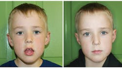
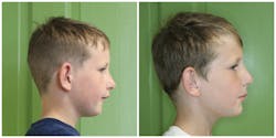
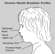
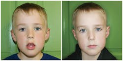
The characteristic appearance of these individuals includes not only a long, narrow face, but often a short upper lip, recessed chin, high and narrow maxilla and forward head posture. This facial appearance at any age is a red flag to which we need to pay attention. The majority of these cases began with airway issues in early childhood. Long before they grew into adults with snoring and sleep apnea, they were the children who suffered from chronic nasal allergies or sinusitis, or whose tonsils and adenoids were chronically enlarged during the years when their teeth and faces were developing. You may have treated these children for years, making notes in the chart year after year about their large tonsils or their gingival inflammation from mouth breathing. You may have referred them to the orthodontist or even the ENT specialist. But was it enough? Did the medical or surgical intervention by the ENT specialist or the orthodontic treatment do enough to reverse the negative esthetic appearance of the long face?
Beyond the referrals to the orthodontist and the ENT specialist, there is still more that we as hygienists can do. Pursue training in orofacial myology/myofunctional therapy. Learning how to help patients reestablish normal ORP is a key piece of the OSA puzzle. (6) Not only would you add a valuable service to your office, but you can help improve the health and wellbeing in the lives of many patients. A proper assessment of facial form and oral muscle function can and should be a routine part of the hygienist’s examination. In fact, assessment and treatment of orofacial myofunctional disorders (OMDs) has been in the ADHA’s Scope of Practice since 1992, yet it is still not taught in most hygiene programs. I would encourage all hygienists to seek out training. It is my hope that someday soon, the recognition and treatment of OMDs will be as routine as assessing periodontal health in our patients.
For the most current dental headlines, click here.
1. Park SY. Sleep Apnea Is a Craniofacial Problem. http://doctorstevenpark.com/sleep-apnea-is-a-craniofacial-problem. Published February 10, 2011. Accessed May 27, 2016.
2. Hanson ML, Mason RM. Orofacial Myology: International Perspectives. 2nd ed. Springfield, IL: Charles C Thomas; 2003.
3. Proffit WR. Equilibrium Theory Revisited: Factors Influencing Position of the Teeth. Angle Orthod. 1978;48(3):175-186.
4. Mason RM, Franklin H. Orofacial Myofunctional Disorders and Otolaryngologists. Otolaryng. 2014; 4:e100. doi: 10.4172/2161-119X.1000e110.
5. Proffit WR. Contemporary Orthodontics. 5th ed. St. Louis: Elsevier Mosby; 2013.
6. Huang YS, Guilleminault C. Pediatric Obstructive Sleep Apnea and the Critical Role of Oral-Facial Growth: Evidences. Front Neurol. 2012;3:184.
7. Valera FC, Trawitzki LV, Anselmo-Lima WT. Myofunctional evaluation after surgery for tonsils hypertrophy and its correlation to breathing pattern: A 2-year-follow up. Int J Pediatr Otorhinolaryngol. 2006;70(2:221-225.


