Extraoral panoramic errors: a summary for dental assistants
Taking radiographs can be tricky, but it doesn't have to be. Here's a thorough guide to successful radiographs every time. For starters, just think of yourself as a photographer and your patient as the model.
This article originally appeared in Dental Assisting & Office Manager Digest. Subscribe to the monthly e-newsletter here.
I often compare a radiographic exposure to a photoshoot session when I talk with our SmarterDA (dental assisting exam prep) users. The cameraperson, or photographer, is the clinician, and the model is the patient. To create the best effect, the photographer (clinician) needs to provide excellent instructions.
Panoramic radiography can be used in the following situations:
• For general surveys of oral health
• To determine the best radiographic supplements for surgical procedures
• For initial and progressive evaluations of orthodontic treatment
• For information about pediatric growth and development
• To review chronological dental eruptions and the axes of permanent tooth eruption
• To evaluate cystic or neoplastic lesions
• To measure the dimensions for implantology
• For historical documentation
• To evaluate the temporomandibular joint, and
• To detect the presence of foreign bodies.(1)
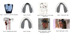
According to the research conducted on positioning errors and published in “Imaging Science in Dentistry,” about 90% of panoramic images were presented with errors while only about 10% were error free. The ranking of positioning errors are as follows: (2)
• Tongue not touching palate during exposure (55.7%)
• Slumped position (35%)
• Patient positioned back (30%)
• Patient positioned forward (18.3%)
• Chin tipped high (17.9%)
• Head turned to one side (17.4%)
• Chin tipped low (16.2%)
• Head tilted to side (12.7%)
• Patient movement during exposure (1.6%)
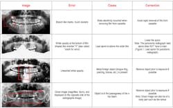
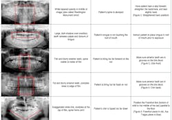
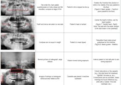
This article summarizes how to detect panoramic radiographic errors, and how to provide instructions about correcting them. The goal is to successfully pass the dental assisting board exams, and also to become the superstar dental assistant everyone wants on their team!
As you can see, small details can make a difference. As a dental professional, it is important to minimize the exposure to radiographs, therefore, avoid retakes.
REFERENCES
1. De Senna BR, Dos Santos Silva VK, França JP, Marques LS, Pereira LJ. Imaging diagnosis of the temporomandibular joint: critical review of indications and new perspectives. Oral Radiol. 2009; 25:86–98.
2. Dhillon M, Raju SM, Verma S, Tomar D, Mohan RS, Lakhanpal M, et al. Positioning errors and quality assessment in panoramic radiography. Imaging Sci Dent. 2012; 42:207–212.
