By Amber Auger, RDH, MPH
The Oral Cancer Foundation reports nearly 50,000 patients will be diagnosed with oral oropharyngeal cancer this year.(1) It is estimated that one person every hour on the hour will die from oral oropharyngeal cancer.(1)Often, oral cancer is discovered after it has metastasized to other areas such as the lymph nodes and neck. Early diagnosis increases the survival rate and decreases the risk of metastasizing; fluorescence technology aids the clinician in detecting abnormal lesions sooner.(2)
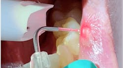
A traditional oral examination relies on reflected light to conduct the exam, whereas tissue fluorescence technology relies on fluorophores that emit their own light at a longer wavelength.(2)The light is blue in color. When directed on healthy tissue, it appears green due to the wavelength technology; when the tissue is abnormal, it appears dark in color.(2) For example, fluorescence can detect changes occurring in the tissue such as cellular and structural changes that are not visible to the eye.(2)
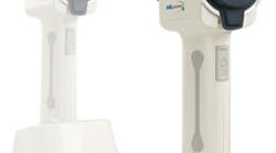
The VELscope Vx (right) uses fluorescence technology and is equipped a phone adapter to capture photos, making referrals to specialists easier. Intended to allow an oral surgeon to identify the area of disease, the VELscope provides vital information needed for surgical excision.(2) Clinical changes can be documented and detected thoroughly with the proper use of the VELscope Vx. This handheld unit is charged with a charging pack and can be easily transported from multiple treatment rooms.
OralID (bottom right) also uses fluorescence technology to allow clinicians to identify abnormal lesions, including those that may be cancerous.(3)The OralID is battery-operated, lightweight, and hand held. When using the OralID chairside, clinicians are instructed to wear OralID eyewear to allow the effects of the blue light to be enhanced during the exam.(3)
When performing the fluorescence assessment, the clinician is looking to detect dark lesions. Once a dark lesion is discovered, a clinical photo should be taken, and the patient should return to the office in two weeks for re-evaluation.(3) The lesion could be dramatic due to temperature trauma or trauma induced by foods. Upon reassessing the patient, if the lesion is present and appears to have darkened, the patient should be referred out to have testing done at the cellular level.(3) This testing will allow the pathologist to determine if the tissue is normal, dysplastic, or malignant and determine the best course of treatment.(3)
The Centers for Disease Control and Prevention recommend the use of the sheath when using the OralID and VELscope.
This technology promotes educational conversations between the patient and clinician. When clinicians use the fluorescence technology, it creates an opportunity to educate our patients about the risk of oral cancer, including tobacco use, alcohol, HPV, lichen planus, betel quid and guta (mostly used in Asia), and weakened immune system.(4) Involving the patient empowers them to reduce their own controllable high risk factors.
Dental hygienists and dentists who consistently perform oral cancer screenings each and every time increase the likelihood of early detection. We are more than prophy providers. We are called to serve our patients through education that will lead to prevention. As dental professionals, our thorough, comprehensive, and well-documented oral abnormality screens should be done on every patient, every time without exception.
References
- Oral Cancer Foundation. Oral Cancer Facts. Available at: http://oralcancerfoundation.org/facts/. Accessed April 12, 2017.
- VELscope. Enhanced Oral Assessment System. Available at: http://www.velscope.com/velscope-products/velscope. Accessed April 13, 2017.
- OralID. Say “No thanks!” to Expensive Devices & Per-Patient Costs! Available at: http://www.forwardscience.com/oralid. Accessed April 13, 2017.
- American Cancer Society. Causes, Risk Factors, and Prevention. Available at: https://www.cancer.org/cancer/oral-cavity-and-oropharyngeal-cancer/causes-risks-prevention/risk-factors.html. Accessed April 13, 2017.


