Incorporating AI into dental school curriculum
The UCLA School of Dentistry has integrated an artificial intelligence (AI) component into its radiographic interpretation training. AI has led to an overhaul of many aspects of the school’s curriculum. It’s become effective not only as a teaching tool but also as an introduction to a technology that students are sure to encounter in their professional lives.
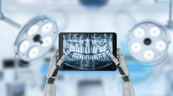
The system chosen is Second Opinion from the California-based dental AI company Pearl. An FDA-cleared cloud-based radiologic AI software platform, Second Opinion instantly analyzes and annotates radiographic dental images, identifying and, where appropriate, quantifying most of the pathologies that are identifiable in x-rays.
AI in dentistry goes hand in hand with digital radiography, which first appeared three decades ago and has become increasingly prevalent, particularly among DSOs. The advantages of digital imaging include greatly reduced radiation exposure, superior image sharpness and dynamic range, and immediate availability of the image.
Dental radiographs have always been subject to a range of interpretations, depending on factors such as ambient illumination, monitor quality, and above all, the skill of the reader. These problems were greater when the radiographic medium was film, and partly as a result of the shortcomings of x-ray film, dental medicine has historically struggled to achieve a perfect diagnostic consistency.
In the early days of digital imaging, its far-reaching potential may not have been obvious to practitioners or patients, for whom having x-rays taken remained as uncomfortable as before. But digital imaging has turned out to be much more than a superior version of film. As in other fields with digital technologies, new possibilities have opened up. We did not foresee how digital imagery, collected as "big data," might affect dental medicine.
That did not become apparent until AI. Trained with large volumes of existing radiographs annotated by practitioners, systems like Second Opinion match the diagnostic accuracy of human radiologists. With AI, digital radiography becomes more than just a superior alternative to film. It’s a complete system that generates and interprets high-quality images and offers a consistent touchstone of accurate diagnosis.
The results of Second Opinion
To evaluate the usefulness of Second Opinion in the curriculum, we compared its performance in caries detection with that of 86 students. The closed-book test consisted of 100 cases from which each student was randomly assigned 10. Seven correct identifications were required to pass. The gold standard of correctness was the consensus of five expert faculty members. In borderline instances, for example regarding the categorization of penetration of caries (E1, E2, etc.), students were credited with a correct answer for either the higher or the lower alternative.
Of the 86, 44 students passed on the first test and 33 passed on a retest. One student required three retests to achieve a passing score of 70%. Second Opinion scored 94%, and only three students did as well or better.
This result underscores the potential utility of Second Opinion as a teaching tool. Providing a granular analysis of radiographs with a high degree of accuracy and reliability, it supplies each student with a faculty member with whom to compare interpretations. AI not only increases the amount of expert feedback available to students, but also reduces faculty workload. “Longitudinal learning" in the form of continuous availability of a high diagnostic standard helps overcome students’ tendency to neglect their diagnostic skills in favor of their procedural ones.
An important aspect of exposure to AI as a teaching tool is that it familiarizes students with the system so they learn how to get the most out of it. However, we find that we have to guard against automation bias. Like any adopters of a new technology, students tend to fall in love with it. They accept its output uncritically.
To assess the influence of automation bias and guard against passive acceptance of computer-generated information, we’re planning studies where original radiographs will be presented alongside AI-annotated versions, some of which will be doctored with false positive or negative diagnoses. The purpose of these anti-tests is to ensure that students continue to develop and use their own judgment in the face of Second Opinion's graphically persuasive and generally accurate analyses.
AI interpretation of dental radiographs is already widely accepted in dental practices, particularly in DSOs, where one of its benefits is to support diagnostic consistency among large numbers of practitioners. IT managers in large organizations proceed with caution, and adoption in schools may be slower than in practices. At UCLA, Second Opinion is not systemwide, with its use limited to teaching and research.
We look forward to having this technology incorporated into our patient care workflow. In academic centers and hospitals, implementations will be reviewed by the IT risk assessment team, a stringent and often lengthy process that’s well worth the wait and effort. In dental medicine as in many other fields, AI is the future. Schools must prepare their students to meet it.
Sanjay Mallya, BDS, MDS, PhD, is a professor and chair of the section of oral and maxillofacial radiology in the Division of Diagnostic and Surgical Sciences at the UCLA School of Dentistry. He is a board-certified oral and maxillofacial radiologist. His research focuses on the molecular mechanisms of oral cancer and parathyroid neoplasia.
About the Author

Sanjay Mallya, BDS, MDS, PhD
Sanjay Mallya, BDS, MDS, PhD, is a professor and chair of the section of oral and maxillofacial radiology in the Division of Diagnostic and Surgical Sciences at the UCLA School of Dentistry. He is a board-certified oral and maxillofacial radiologist. His research focuses on the molecular mechanisms of oral cancer and parathyroid neoplasia.
