By Maria Perno Goldie, RDH, MS
In 2001, the NIH had a Consensus Development Conference, the first sponsored by the NIH on dental caries, which provided an excellent venue to describe the success that has been achieved in reducing caries prevalence to date.(1) They confirmed that effective preventive practices, such as the use of fluoride, sugarless products, and dental sealants were still useful and suggested clinical studies to identify more conservative and effective nonsurgical and surgical approaches to controlling the caries process. They concluded that current diagnostic practices (in 2001) were inadequate to achieve the next level of caries management in which noncavitated lesions are identified early so that they can be managed by nonsurgical methods.In the case of dental decay, it has traditionally been diagnosed with visual inspection, the use of an explorer, and traditional radiographs. The 2001 NIH panel supported the use of visual and tactile techniques for detecting tooth decay; however, the use of an explorer to detect occlusal decay was not endorsed.(1)Explorers often present incomplete or inaccurate data and may actually break and intact surface layer. This did not, and does not, stop clinicians from using explorers in this fashion. The consensus was that dental caries is an infectious disease, is communicable, and in order to prevent tooth decay the caries process must be controlled. Restorations have no effect on the caries process and may actually cause greater risk of disease. In 2005, the debate concerning explorers continued. Some said yes, an explorer is a time-tested tool for carious lesion detection.(2) Probing for caries is part of the standard of care when examining teeth during a dental examination, and this procedure is well-accepted by dentists, dental insurance companies and patients. A 1972 document is the basis for the above, which defines that “[an] area is carious when the explorer ‘catches’ or resists removal after the insertion into a pit or fissure with moderate to firm pressure, and when this is accompanied by one or more of the following signs of caries: “a softness at the base of the area; “opacity adjacent to the pit or fissure; “softened enamel adjacent to the pit or fissure.”(3)The counter point to that point of view is that use of an explorer can lead to misdiagnosis and disrupt remineralization.(2) The results of several studies indicated that use of the dental explorer was of limited value for the detection of occlusal caries.(4) One study concluded that there is strong evidence to support the elimination of the use of the dental explorer in the historical manner.(5) The explorer will stick in a very cavitated lesion, but it will give no indication in early stages of decalcification, when the tooth could potentially be remineralized. There is also apprehension that the explorer may inoculate healthy areas with bacteria transferred from another tooth. Today, the goal of the examination should include the identification of lesions or demineralized areas at the precavitation stage that may be reversed or arrested if the thin surface layer covering the demineralized area remains intact. Fast forward to 2011. We are becoming more sophisticated and distinguishing between dental caries and dental decay, and between caries diagnosis and caries detection. Diagnosis is the art or act of identifying a disease from its signs and symptoms.(6) This is distinct from the detection of the signs and symptoms themselves. The diagnosis forms the basis for making informed treatment decisions. The art and science of diagnosis is based on the hypothesis that diseases can be distinguished from their signs and symptoms. Innovations in caries detection technology make it easier for early diagnosis and preventive treatment to be the standard of care.This issue will address some of the new technologies available. Laser fluorescence, LED fluorescence, LED fluorescence with intraoral camera technology, and digital radiography are a few of the technologies available to us today.
ACTEON’s SOPROLIFE (light-induced fluorescence evaluator) is an intraoral camera and caries-detection device in one. Using autofluorescence technology, lighting switches from 4 white LEDs to 4 blue LEDs enabling the clinician to see variations in the tooth’s state of dentin health. It has been shown to assist in caries detection and treatment.(7) SOPROLIFE captures images in three modes (portrait, intraoral, and macro), while facilitating detection of the occlusal and interproximal caries frequently missed by traditional X-rays.(8) www.acteongroup-products.com/SOPROLIFE
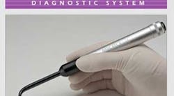
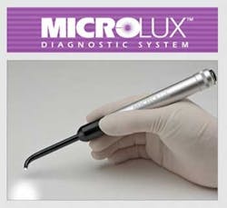
Spectra Caries Detection Aid supports a minimally invasive treatment approach and early caries detection. Treatment options are clear with color images, and it complements radiographs during patient exams. Images can be captured, stored, compared and retained. SPECTRA detects tooth decay by measuring increased light-induced fluorescence. www.airtechniques.com/dental/caries_detection_aid.cfm The Midwest Caries I.D. is LED Fluorescence, or a LED light aimed at the tooth. The unit measures the reflection and refraction of this light for the unique optical signatures of healthy or decalcified enamel. The Midwest Caries I.D. has demonstrated that occlusal caries diagnosis can be improved by LED diagnosis compared to radiographic assessment. With respect to approximal lesions, decay is detectable with the probe positioned on the marginal ridge of the occlusal surface. However, further in vitro and in vivo studies have to evaluate the possibility of reducing or even replacing radiographic examinations.(9,10) The Midwest Caries I.D.™ technology emits a soft (LED) light and captures the resulting reflection and refraction in the tooth. A specific optic signature identifies the presence of caries. Healthy tooth structure is generally more translucent than a decalcified one; consequently, it has a different optic signature. The light reflected from the decalcification of the enamel is captured by fiber optics and is converted to electrical signals for analysis. www.cariesid.com/The DIAGNOdent from KaVo has been on the market the longest period of time. Laser fluorescence, or laser light of a specific frequency is directed at the tooth, and the amount of reflected light is measured. Healthy teeth exhibit little to no fluorescence, but carious tooth structures exhibit fluorescence proportionate to the degree of caries. The diagnostic accuracy of DIAGNOdent was significantly better than that of radiography (p< or =0.001). In this in vitro study of detection of occlusal caries, the diagnostic performance of the DIAGNOdent method was superior to that of radiography.(11) Research studies can be found on page 6: www.drfrankgodino.com/_media/pdf/61_Brochure_diagnodent.pdfThe CarieScan PRO is an early caries detection system and its intended use is: "For use by dental professionals as an aid in the diagnosis and monitoring of dental caries." (12) The CarieScan PRO is a device for the detection and monitoring of caries by the application and analysis of ac impedance spectroscopy (ACIST). The PRO uses a single-use disposable sensor. The sensor is held against the tooth being examined and the detection process takes about four seconds to complete. The result is displayed on the LCD screen and the color LED display. The electrical current used is extremely low and cannot be felt by the patient. The system is battery operated. It enables dental professionals to evaluate decay in teeth using ACIST by providing information about whether the tissue is healthy, in the early stages of decay or already significantly decayed. The CarieScan PRO should not be used on patients with cardiac pacemakers. us.cariescan.com/CariScreen (Oral BioTech) evaluates caries risk is by measuring oral bacteria load using ATP Bioluminescence. A swab collects plaque from the teeth, and a meter measures the reaction with bioluminescence reagents within the swab to provide a numerical score. www.carifree.com/dentists/products/cariscreen.phpMicrolux Transilluminator uses fiber optic transillumination. A fiber optic-focused LED is directed through the tooth where decay is suspected and areas of decay appear darker as the demineralized tooth structure scatters and absorbs more light than healthy tissue. www.addent.com/prod-microlux1.htmlDigital Radiography with Software Analysis are taken and analyzed by the software. A set of data-derived algorithms is used to classify and determine the severity of proximal caries. An example is LOGICON Caries Detector software, Kodak Dental Systems.(13)www.carestreamdental.com/en/digital-imaging/software-and-accessories/logicon-caries-detector.aspx
References
1. Diagnosis and Management of Dental Caries Throughout Life. NIH Consensus Statement 2001 March 26–28; 18(1) 1–30. 2. Hamilton JC and Stookey G. Should a dental explorer be used to probe suspected carious lesions? J Am DentAssoc 2005; 136; 1526-1532.3. Radike AW. Criteria for diagnosis of dental caries. In: Proceedings of the Conference on the Clinical Testing of Cariostatic Agents, American Dental Association, Chicago, Illinois, October 14-16, 1968. Chicago: ADA Council on Dental Research; 1972:87-8. 4. Lussi A. Validity of diagnostic and treatment decisions of fissure caries. Caries Res 1991; 25:296-303.5. Anusavice K. The maze of treatment decisions. In: Fejerskov O, Kidd EA, eds. Dental caries. The disease and its clinical management. Malden, Mass.: Blackwell; 2003:251-65. 6. Nyvad B. Diagnosis versus Detection of Caries. Caries Res 2004; 38:192–198.7. Terrer E, Koubi S, Dionne A, et al. A new concept in restorative dentistry: light-induced fluorescence evaluator for diagnosis and treatment. Part 1: Diagnosis and treatment of initial occlusal caries. J Contemp Dent Pract. 009;10:E086-E094.8. Terrer E, Raskin A, Koubi S, et al. A new concept in restorative dentistry: LIFEDT-light-induced fluorescence evaluator for diagnosis and treatment: part 2 - treatment of dentinal caries. J Contemp Dent Pract. 2010; 11:E095-E102. 9. F. Krause, D.J. Meiner, R. Stawirej, S. Jepsen, A. Braun. LED Based Occlusal and Approximal Caries Detection In Vitro. Abstract 0142, IADR, 2008.10. F. Krause, D.J. Melner, R. Stawirej, S. Jepsen, and A. Braun. Led Based Occlusal And Approximal Caries Detection In Vitro. Abstract 0526, IADR, 2008. 11. Shi XQ, Welander U, Angmar-Månsson B. Occlusal caries detection with KaVo DIAGNOdent® and radiography: an in vitro comparison. Caries Res. 2000 Mar-Apr;34(2):151-8.12. us.cariescan.com/products-2/cariescan-pro13. Gakenheimer, David C., "The Efficacy of a Computerized Caries Detector in Intraoral Digital Radiography." Journal of the American Dental Association 133 (2002): 883-890.Additional Reading
Zandoná AF and Zero DT. Diagnostic tools for early caries detection. J Am Dent Assoc 2006 137(12): 1675-1684.Zero DT, Fontana M, Martínez-Mier EA, Ferreira-Zandoná A, Ando M, González-Cabezas C, and Bayne S. The Biology, Prevention, Diagnosis and Treatment of Dental Caries: Scientific Advances in the United States.. J Am Dent Assoc 2009 140(suppl_1): 25S-34S.Barbería E, Maroto M, Arenas M, and Silva CC. A Clinical Study of Caries Diagnosis With a Laser Fluorescence System. J Am Dent Assoc 2008 139(5): 572-579.de A. Rodrigues J, Hug I, Diniz MB, Cordeiro RCL, and Lussi A. The Influence of Zero-Value Subtraction on the Performance of Two Laser Fluorescence Devices for Detecting Occlusal Caries In Vitro. J Am Dent Assoc 2008 139(8): 1105-1112.
Maria Perno Goldie, RDH, MS


