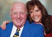
A Daughter’s New Smile Leads Dad His Own Fountain of Youth
How many times have you given a treatment presentation, only to find out months later that your technical jargon went right over the patient’s head? Not only did it go over the patient’s head, it went right down the street to another provider. Rehabilitating a smile, although often complicated, first requires a ready and willing patient.
Keeping the treatment options and initial patient experience simple has proven worthy of careful consideration. Simple options for investigating the possibilities of smile enhancement have also opened the door to increased staff involvement as well as increased case acceptance. Below is a case study of a father and daughter treatment plan that was a great success using these techniques.
Comprehensive analysis of every case prior to treatment is an absolute must. A comprehensive examination should include a smile analysis,1,2,3,4,5 full-mouth series of radiographs,6 models, jaw records, and bite analysis.7,8 Often, a patient’s budget becomes a major factor in treatment. Thorough records allow success in phasing treatment. Knowing the status of the patient prior to treatment and having a clear end goal in mind allow a higher degree of success in the oral rehabilitation of form, function, and esthetics.9
In our world with heightened emphasis on physical appearance, a series of dental photographs10 is often included in initial record-taking. As they say, time is money. Both can drive treatment for the doctor and patient. Patients may not have time today for initial records, and the doctor may not have time to slip a comprehensive examination into an already busy day if it is not prescheduled. Patients may find themselves late for their next appointment. In this fast-paced world of email and pagers, doctors as well as patients may be more accepting of a quicker, simpler approach to treatment options.
Simple imaging
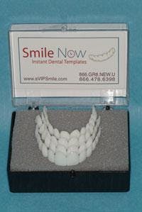
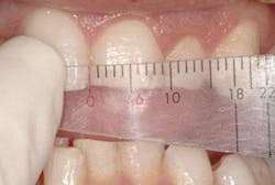
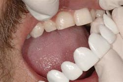
Simple can mean different things to different people. For years, I had used quick resin mockup directly in the patient’s mouth as a “simple” way for all to visualize the possibilities for cosmetic dentistry. With the advent of digital imaging, the use of computer enhancement as a visual aid to investigate the possibilities of smile design has also been helpful. However, it is time-consuming to take photographs, download them, and enhance them to consider thepossibilities of a new smile. Instead, I have attempted a newer technique using an instant dental template 11 (Figure 1). Within one minute, you can show patients what a beautiful new smile can look like. The template is currently available in four sizes. The existing width of the central incisor is measured and the corresponding template chosen. The template can be slipped in over the existing teeth and held in place, or it can be relined with a temporary material for view without fingers in place. It’s quick and simple for auxiliary use and ideal for increasing case acceptance.
The daughter arrives at our doorstepGetting staff involved
Figure 1 - Smile-Now instant dental templates
We were in the office on a Thursday afternoon, wrapping up for the day, when a new patient walked in the door. She stated she had seen a local ad for cosmetic dentistry and wanted to see if she was a candidate. The clinical assistant asked if she had a moment. She did and with the simple technology described above - in an instant - the dental template for her corresponding 8 mm central was tried in (Figure 2) by the clinical assistant. Having had esthetic treatment herself, she was able to discuss treatment as well as options from a team member’s perspective. This noninvasive technique of a simple dental template demonstration allows the team to feel more a part of the treatment process. The prospective patient was amazed! She wanted to get started right away. Now, an appointment could be scheduled for records. No longer are records taken without a commitment from the patient to proceed with treatment.
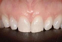
The following week, models, photos, and jaw measurements were taken so that records could be placed on an adjustable articulator and a diagnostic wax-up could be completed. Treatment recommendations included in-office teeth whitening to establish the desired shade, Empress porcelain restorations on all upper teeth, and replacement of old crowns in the lower as well as Empress on the lower anterior centrals to replace old bonding and correct malalignment (Figure 3). Daughter’s enthusiasm leads to treatment plan for Dadnull
Increasing case acceptance
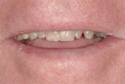
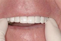
Teh patient began treatment immediately by scheduling a Brite-Smile in-office whitening appointment. Although porcelains were advised for the entire upper arch and the lower two front teeth, finances were a concern. The patient could not afford all recommendations. Through the diagnostic wax-up process, I was able to segmentally treat the patient. Goals included using the Waterlase to recontour gums, followed by Empress porcelain restorations from maxillary first premolar to premolar. Although the importance of the buccal corridor was emphasized to the patient, it was decided that the maxillary molars would be treated in the near future. In addition, subsequent treatment would also include mandibular central veneers and replacement of old mandibular crowns. Routine treatment was rendered; however, on the day of provisionalization, the patient was so excited that she brought her father in to investigate the benefits of smile enhancement for him (Figure 4). Once again, simple templates allowed team members to discuss cosmetic dentistry with Dad (Figure 5). Simple discussion led to immediate case acceptance (Figure 6). Unfortunately, it was Dec. 23, 2004. All of us working mothers were preparing a Christmas feast the following day! Smile enhancement for Dad would have to remain a vision until 2005. Appointments were scheduled for records to turn this exciting vision into reality.
Treatment
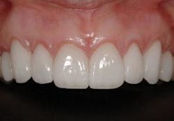
By the time Dad started, daughter was completing Phase I of her new smile. Empress-layered porcelains were tried in with try-in paste for patient approval. Anesthesia was obtained with Articaine 4 percent HCL with 1:100,000 epinephrine to cross the buccal plate of bone and offer comfort without the need for palatal injections. A small Ultradent “00” cord was placed for retraction to obtain an ideal seal and minimize microleakage of the resin cement. Porcelains were cemented under rubber dam isolation after debriding with Hibiclens, a chlorhexidine gluconate, as a cleansing agent. Teeth were conditioned with Ultradent Ultra-etch 35 percent phosphoric acid for 10 to 15 seconds and rinsed. Care was taken during the etchant process not to over-etch.12 Superseal wetting agent (Phoenix Dental) was applied and lightly air-dried so as not to leave teeth overdessicated or puddled with fluid. Kerr’s OptiBond Solo Plus universal dental adhesive was applied with a brush and dispersed with air syringe alone and cured. A hand syringe has been dedicated to air alone in each treatment room for this purpose. A second layer was applied by the doctor as the assistant applied a thin coat on the internal surface of the porcelains. Next, air dry with air syringe once again and light cure. Porcelains were loaded with Rely-X from 3M/ESPE. The centrals were seated first, followed by laterals, cuspids, and so on. Once seated, excess resin was removed with a brush and the tacking process - five seconds tack per tooth - began. Once tacked in place, the margins were checked, and the final cure completed. A Proxi-disc by Centrix was used to separate the contacts followed by floss. Glycerine was applied to the margins and cured to provide an air-inhibited layer to minimize microleakage. The rubber dam was now removed and digital radiographs were taken to verify seat and check for excess cement flash (Figure 7). Occlusion was checked and balanced to offer even, centric contacts and a smooth anterior guidance sliding up the cuspids with a smooth transition in cross-over to the central incisors.
Now it’s Dad’s turn Dad’s differential diagnosis included gum recession; left cross-bite; asymmetrical gum heights; old, leaking amalgams in the left premolar region; and caries under existing crowns. The patient did not wish to undergo periodontal grafting to correct the gum recession; however, the upper lip line masked the asymmetry of the gumline nicely. In addition, the lipline worked to the author’s advantage in that both maxillary centrals were nonvital with an old and possibly irretrievable post in the upper left central under the existing crown.null
null
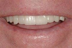
The author was able to correct the cross-bite through porcelain coverage. In addition, anterior guidance was maintained13 and the envelope of function14 was opened up in an attempt to minimize further recession in the upper anterior segment. The day of beauty was here (Figure 8). Upon removing the provisional, the upper right central fractured. An in-office post was fitted and the crown was placed over the prefabricated and cemented post with core past. This was not ideal, but certainly functional. Due to the low lipline, the author felt that this course of treatment, although not ideal, could function nicely as the final restoration. As you can see from the smile photographs, unless the lip is retracted, shade variation from one central to the other is not visible. Cementation occurred under rubber dam isolation. Hibiclens was used to debride the preparations after the temporaries were removed. Steps as noted above in the daughter’s case were followed for father also. Everyone’s a winner Nothing is better than a happy patient. And in this case, two happy patients are sharing their dental experiences and success (Figure 9).
Simplification of treatment options and the presentation process can make everyone a winner. Patients and team can visualize the benefits much faster. In addition, the team experiences the benefits of increased involvement in dental treatment. This translates into increased case acceptance and, in this specific situation, a multigeneration makeover! Father and daughter are both smiling. Everyone - doctor, team, and patients - experiences the joy of smile design and dental rehabilitation.null
null
null
References
1 Eubank J, Morley J. Macroesthetic elements of smile design. JADA 2001; 132.
2 Ahmad I. Geometric considerations in anterior dental aesthetics: restorative principles. Pract Periodont Aesthet Dent 1998; 10(7):813-822. 3 Hunt K. Full-mouth multidisciplinary restoration using the biological approach: a case report. Pract Procedural Aesthet Dent 2001; 13(5): 399-406. 4 Lichter J, Sauco M, et al. What’s behind your smile? Journal of Michigan Dental Assoc Nov. 2002. 5 American Academy of Cosmetic Dentistry. A Guide to Accreditation Criteria. 6 Updegrave WJ. Right angle dental radiography described, simplified, and standardized, part II. Instrumentation. L.D. Pankey Institute for Advanced Dental Education, Continuing Education 1982; Article #4, III(1):39. 7 L.D. Pankey Institute, Continuum Level I, Accuracy in Articulation. Level I manual, section 3:2-10. 8 Bryant W. L.D. Pankey Institute Level 4, Philosophy of Occlusal Rehabilitation, section 2:43-56. 9 Soll J. Step into action. Dentistry Today Sept. 1998. 10 American Academy of Cosmetic Dentistry. A Guide to Accreditation Photography. 11 Smile-Now, Instant Dental Templates. www.yoursmilenow.com, Presented as new product at the American Academy of Cosmetic Dentistry Annual Scientific Session 2005, Nashville, Tenn. 12 Poss S. Advanced Anterior Esthetics (lecture). Sponsored by Expertec Dental Laboratory, Inc., Westland, Mich., Feb. 4, 2005. 13 Wynn W. Considerations for establishing and maintaining proper occlusion in the aesthetic zone. Dentistry Today April 2004. 14 Bryant WE. An approach to the study of occlusion through an understanding of anterior control. L.D. Pankey Institute C-3 Manual, section-2:3-17.Mary Sue Stonisch, DDS, FAG
Dr. Stonisch maintains a Smile Enhancement Studio in Grosse Point, Mich., limited to comprehensive restorative care and esthetic dentistry. She is an accredited member of AACD and a fellow of the AGD and the American Academy of Dental Facial Esthetics. You may contact her at [email protected] or www.smileenhancement studio.com.

