Oral pathology case: Detecting a life-threatening disease
Presentation and chief complaint
A 77-year-old female presents to the office for a new-patient exam. Her chief complaint was that she was due for a cleaning and check-up.
Health history
Health history included cholesterol and blood thinner medications, type 2 diabetes, history of chronic obstructive pulmonary disease (COPD), high blood pressure, and sensitivity to penicillin.
Clinical exam
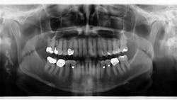
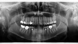
Assessment of the panoramic radiograph revealed two corrugated, irregularly shaped radiopaque masses on the right posterior angle of the mandible, each measuring approximately one inch in length. The area was not tender to palpation, and the patient had no knowledge that the lesions were there.
Differentials
- Calcified carotid artery/arteriosclerosis
- Calcified lymph nodes
- Sialoliths
- Phleboliths
- Calcification of the styloid and stylomandibular ligaments
- Calcification of the triticeal, cricoid, and thyroid cartilages
**See image below**1
Final diagnosis
Calcified carotid artery/arteriosclerosis:
“Atherosclerosis is a chronic inflammatory disease of multifactorial origin, characterized by artery walls thickening or elasticity loss, many times associated with the presence of atheromas.”2 As physicians in the specialized field of dentistry, we often find ourselves in a unique position to detect these lesions (or a differential), due to the frequent utilization of panoramic radiography. Observation of the first cervical vertebra area is a vital diagnostic aid in the early diagnosis of calcification of carotid arteries.3 Why is this important? “The presence of atheromas in the carotid arteries of neurologically asymptomatic individuals is frequently associated with future development of cerebrovascular diseases.”2 Simply put—we can save lives.
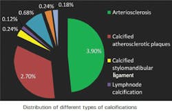
The frequency of the differentials, as mentioned earlier, is noted in the graph below.4 While calcification in the arteries and lymph nodes is prevalent, a definitive diagnosis is vital.
While panoramic radiography is often the genesis for referral and further testing, it is not indicated as a sole diagnostic tool. A referral to an ENT or internal medicine physician is the typical protocol one should follow, which generates the order for ultrasonic imaging of the area in question. “Carotid ultrasonography is the noninvasive imaging method utilized as the gold standard in the diagnosis of carotid artery calcification.”2
This particular patient was referred to an oral surgeon for assessment. The oral surgeon concurred with the initial thought that this was a carotid artery calcification. The patient was then referred to an ENT for an ultrasound of the area, at which time the final diagnosis was given. It was concluded that the lesions were not large enough to hinder blood flow so as to recommend invasive treatment, such as stent placement. The patient has been placed on a recall program for continuous assessment and monitoring.
Editor's note: Originally published February 19, 2018. Updated September 2024.
References
- Carter LC. Soft tissue calcifications and ossifications. Pocket Dentistry website. https://pocketdentistry.com/28-soft-tissue-calcifications-and-ossifications/. Accessed February 19, 2018.
- Brasileiro Junior VL, Luna AHB, Sales MAO, Rodrigues TLC, Sarmento PLFA, Mello Junior CF. Reliability of digital panoramic radiography in the diagnosis of carotid artery calcifications. Radiol Bras. 2014;47(1):28-32. doi: 10.1590/S0100-39842014000100011.
- Nilton A, Deana NF, Garay I. Detection of common carotid artery calcifications on panoramic radiographs: prevalence and reliability. Int J Clin Exp Med. 2014;7(8):1931-1939.
- Vengalath J, Puttabuddi JH, Rajkumar B, Shivakumar GC. Prevalence of soft tissue calcifications on digital panoramic radiographs: A retrospective study. J Indian Acad Oral Med Radiol. 2014;26(4):385-389. doi: 10.4103/0972-1363.155676.
About the Author
Stacey L. Gividen, DDS
Stacey L. Gividen, DDS, a graduate of Marquette University School of Dentistry, is in private practice in Montana. She is a guest lecturer at the University of Montana in the Anatomy and Physiology Department. Dr. Gividen has contributed to DentistryIQ, Perio-Implant Advisory, and Dental Economics. You may contact her at [email protected].


