Editor's note: Originally published April 23, 2018. Formatting updated November 15, 2022.
Presentation and chief complaint
A healthy 54-year-old female presents for her routine dental exam and cleaning. Her chief complaint at this visit is that she has a lump under her tongue on the left side that has been there for approximately two weeks. The patient commented that it hurts when she eats because the food irritates that spot directly. Furthermore, the lump fluctuates in size to a small degree, but it never really goes away.
Clinical exam
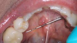
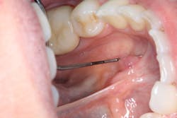
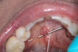
Clinical assessment reveals a red, tissue-colored, raised, fluctuant mass on the left side of the lingual frenum, in the area of the submandibular gland duct. There is a slight tenderness to palpation with some firmness to the lesion overall. The mass measures approximately 4 mm x 5 mm in length (see photos below).
Differentials
- Mucocele
- Mucus retention cyst
- Sialolith
Definitive diagnosis
Mucus retention cyst
Discussion
A mucus retention cyst is defined as a “swelling caused by an obstruction of a salivary gland excretory duct resulting in an epithelial-lined cavity containing mucus."1 The mucus-filled cavity is lined with epithelium,2 but rarely involves the major salivary glands.1
Mucus retention cysts can be identified by the following characteristics:
- They can occur at any age, but primarily in adults who are in their third to eighth decades of life.1
- If found in the major salivary glands, 90% are usually in the parotid gland and 10% are in the submandibular and sublingual glands.1
- The cysts can resemble mucoceles and low-grade mucoepidermoid carcinomas.
- The most common sites of occurrence are the floor of the oral cavity, followed by the buccal mucosa, and the lower lip.1
- Presentation is as a fluctuant, painless, self-contained superficial mass.
- Treatment is by simple excision, and sometimes the lesions are self-limiting.
- Treatment and ongoing assessment
In this particular case, the patient was instructed to leave the lesion alone to see how it would manifest over the course of the next two weeks. A follow-up call revealed that the lesion had indeed receded and was, for the most part, gone. Assessment for any changes or questionable pathology in this area will be noted at future recare exams.
References
- Sapp JP, Eversole LR, Wysocki GP. Contemporary Oral and Maxillofacial Pathology. St. Louis: MO: Mosby; 1997.
- Wood NK, Goaz PW. Differential Diagnosis of Oral and Maxillofacial Lesions. 5th ed. St. Louis, MO: Mosby; 1997.
About the Author
Stacey L. Gividen, DDS
Stacey L. Gividen, DDS, a graduate of Marquette University School of Dentistry, is in private practice in Montana. She is a guest lecturer at the University of Montana in the Anatomy and Physiology Department. Dr. Gividen has contributed to DentistryIQ, Perio-Implant Advisory, and Dental Economics. You may contact her at [email protected].


