Pathology case: This lesion’s unknown etiology put it into a high-risk category
Editor's note: Originally published May 2017. Updated July 2024.
Case presentation
A 77-year-old male presented for his annual checkup and exam. He visits our office only once a year and alternates with his dentist in California the other six months, where he is a snowbird during the winter.
The patient's chief complaint was a white spot on the left side of his tongue that had been present for just less than a year.
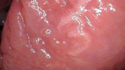
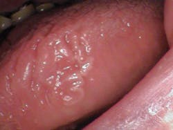
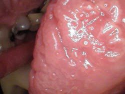
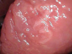
Clinical assessment revealed a 6 mm x 8 mm white leukoplakic lesion on the left lateral border of the tongue. The lesion was not painful to palpation or touch, and it was slightly corrugated with irregular borders.
The patient's health history was noncontributory except for blood pressure medication.
Differential diagnoses
- Squamous cell carcinoma
- Acute glossitis with squamous hyperplasia
- Localized lichen planus
Definitive diagnosis
Acute glossitis with superficial erosions superimposed on pseudoepitheliomatous squamous hyperplasia with mild atypia
Discussion
The unknown etiology of the lesion automatically put it into a category that made it a high-risk leukoplakia, especially since it had been present for just under a year. The patient was referred to an oral surgeon for excisional biopsy. The final diagnosis, as given above, did not reveal any evidence of malignancy. Because the findings were consistent with inflammatory changes, it was recommended that the patient be monitored for any future premalignant changes or tendencies. A three-month follow-up was recommended.
I previously presented a leukoplakic pathology case. The following is taken from that case and gives a general background on leukoplakic lesions and is a good reference to follow.
The presence of leukoplakic lesions in the oral cavity is always cause for evaluation and follow-up. The World Health Organization defines leukoplakia as “a white patch or plaque that cannot be characterized clinically or pathologically as any other disease.”1
Leukoplakic lesions are one of the more common forms of epithelial dysplasia, typically discovered during routine exams; they are usually asymptomatic and represent 6.2% of all oral biopsy specimens.1 Leukoplakia and squamous cell carcinoma (SCC) share many of the same etiology factors,1 and approximately 5.4% of leukoplakic lesions become SCC.2 It is, therefore, imperative that the following be considered1:
- Assess etiologic factors (table 1)
- Categorize the lesion as a high- or low-risk specimen (table 2)
- Assess differentials (table 3)
- If warranted, biopsy the lesion for a definitive diagnosis
Table 1: Etiologic factors of leukoplakia
- Tobacco products
- Ethanol
- Hot, cold, spicy, and acidic foods and beverages
- Alcoholic mouth rinse
- Occlusal trauma
- Sharp edges of prostheses or teeth
- Actinic radiation
- Syphilis
- Presence of Candida albicans
- Presence of viruses
Table 2: High-risk leukoplakia
- Red component
- Raised component
- Presence in high-risk oval
- Tobacco and alcohol use
- Nonsmoker and unknown etiology of lesion
- Nonreversible type
- Microscopic atypia
Table 3: Differential diagnosis of homogeneous leukoplakia
- Lichen planus
- Leukoedema
- Cheek-biting lesion
- Smokeless tobacco lesion
- Lupus erythematosus
- Hyperplastic or hypertrophic candidiasis
- Hairy leukoplakia
- Electrogalvanic or mercury contact allergy
- Verrucous or squamous cell carcinoma
- Verruca vulgaris
- White sponge nevus
References
- Wood NK, Goaz PW. Differential Diagnosis of Oral and Maxillofacial Lesions. 5th ed. Mosby; 1997:75-77, 98-103, 106-107.
- Sapp JP, Eversole LR, Wysocki GP. Contemporary Oral and Maxillofacial Pathology. Mosby; 1997
About the Author
Stacey L. Gividen, DDS
Stacey L. Gividen, DDS, a graduate of Marquette University School of Dentistry, is in private practice in Montana. She is a guest lecturer at the University of Montana in the Anatomy and Physiology Department. Dr. Gividen has contributed to DentistryIQ, Perio-Implant Advisory, and Dental Economics. You may contact her at [email protected].


