Editor's note: Originally published February 21, 2019. Formatting updated November 15, 2022.
Presentation and clinical exam
A 28-year-old male presented for routine six-month hygiene recall. Initial information was as follows:
- Noncontributory medical history
- Patient denies tobacco use or alcohol abuse
- No prescription medications
Related reading: When Lemonheads won't work
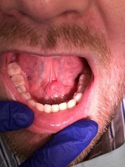
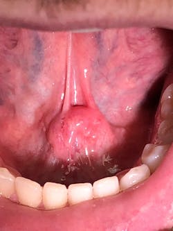
During an oral cancer screening, a soft-tissue lesion was noted on the floor of the mouth at the junction of the ventral surface of the tongue (figure 1).
The lesion was offset to the left, but appeared to cross the midline based upon the position of the frenum. The lesion was about 1.5 cm x 1 cm. Upon palpation, the tissue was firm but not indurated and mobile as one mass. The patient reported no symptoms other than mild discomfort upon palpation (figure 2). When asked about the duration of the lesion, the patient stated he had been aware of it for around two to three weeks, but it was not bothering him.
Differential diagnosis
Differential diagnosis includes:
- Sialolith of the submandibular gland blocking Wharton's duct
- Ranula
- Mucous retention cyst
- Allergic sialadenitis
- Squamous cell carcinoma
Definitive diagnosis
Sialolith of the submandibular gland blocking Wharton's duct
Discussion and treatment
A sialolith is defined as calcified organic matter often found in the draining duct of salivary glands.1 Wharton's duct is a common culprit due to sharp curves, length of duct, and a narrow punctum.2 Treatment modalities are based upon size and severity of the stone. Smaller stones can be treated nonsurgically with dietary changes (increasing sour food intake) with hopes of excretion. Larger stones are typically treated by surgically removing the stone.3
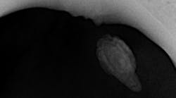
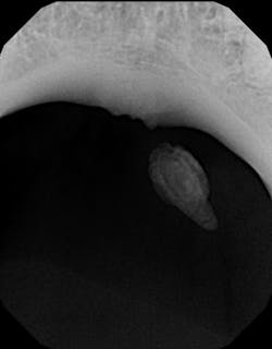
Diagnosis was confirmed with a radiograph of the affected area, showing a calcified stone that approximated the size and location of the lesion (figure 3). Once advised of the treatment options, the patient opted for surgical removal with a local specialist.
References
- Neville BW, Damm DD, Allen CM, Bouquot JE. Oral & Maxillofacial Pathology. 2nd ed. Philadelphia, PA: W.B. Saunders; 2002: 393-395.
- Grases F, Santiago C, Simonet BM, Costa-Bauzá A. Sialolithiasis: mechanism of calculi formation and etiologic factors. Clin Chim Acta. 2003;334(1-2):131-136.
- Wilson KF, Meier JD, Ward PD. Salivary gland disorders. Am Fam Physician. 2014;89(11):882-888.
About the Author
Travis G. Heaton, DDS
Travis G. Heaton, DDS, is a graduate of the University of Texas School of Dentistry at Houston. He maintains a private practice in Tyler, Texas.


