Editor's note: Originally published May 5, 2015. Updated January 2023.
Case presentation
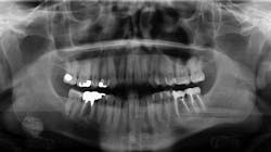
A healthy, 36-year-old male presented for a comprehensive exam. A panoramic radiograph revealed a well-defined, radiopaque lesion approximately 2x2 cm in size in the right submandibular region. The patient was unaware of its existence. The lesion was slightly palpable upon extraoral manipulation, but not necessarily tender.
Diagnosis and treatment
Sialolith of the right submandibular gland
Sialoliths are calcareous (radiopaque) deposits in the ducts of the major or minor salivary glands or within the glands themselves.1 Nearly 80% of all sialoliths affect major salivary glands, the most common being the submandibular gland.1,2 Predilection for the submandibular gland (~73%) and duct may result from gravity and the fact that the oral terminus is superior to the gland.1 Prolonged blockage can lead to degeneration and eventual shutdown of the gland; salivary retention with ductal dilation results in pain and swelling.2 Glands that are no longer functional become subject to retrograde bacterial infections.1 Any swelling is notably present at mealtimes when there is stimulation for salivary production.
Treatments of major gland sialoliths include manual manipulation, surgical “cut-downs” into the duct, or removal.
This particular patient was seen by an oral surgeon and a CT scan was obtained. It was recommended that the lesion be removed; however, since the lesion was asymptomatic at the time, the patient was quite reluctant. The patient is seen on a regular basis with subsequent radiograph evaluation to monitor for changes. The recommendation for removal still stands.
Diagnosis and documenting of pathology is key in all aspects of oral health-care management. What is frustrating from a provider perspective is that if something doesn’t hurt or bother a patient, the inclination to treat it is not there. It is our duty then to respect the patient’s autonomy, document and continue to monitor, and recommend definitive care.
You may also be interested in...
The "non-bothersome" soft-tissue lesion
References
1. Wood NK, Goaz PW. Differential Diagnosis of Oral and Maxillofacial Lesions. 5th ed. Mosby Publishing; 1997:470-472. 2. Sapp JP, Eversole LR, Wysocki GP. Contemporary Oral and Maxillofacial Pathology. Mosby Publishing; 1997:326-328.
About the Author
Stacey L. Gividen, DDS
Stacey L. Gividen, DDS, a graduate of Marquette University School of Dentistry, is in private practice in Montana. She is a guest lecturer at the University of Montana in the Anatomy and Physiology Department. Dr. Gividen has contributed to DentistryIQ, Perio-Implant Advisory, and Dental Economics. You may contact her at [email protected].


