Pathology case: Molars with irregular eruption patterns
Editor's note: Originally published in December 2015. Updated June 2023.
Case presentation
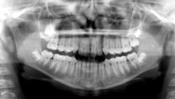
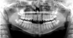
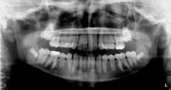
Healthy 11- (figure 1) and 14-year-old (figure 2) males present to the office for new-patient exams. Clinically, it was noted that the eruption patterns were not as they would typically be, primarily for the second molars in the 14-year-old. A panoramic radiograph was taken, revealing large radiopacities around the second molars on the right side. Both patients reported lack of pain upon palpation in these areas.
Diagnosis: Ectopic third molar
Discussion and treatment
According to an article in Contemporary Clinical Dentistry, “Maxillary second molar impaction in the adjacent ectopic third molar: Report of five rare cases,”1 this is not a common finding, with reports of 0.18%, and another study showing a 0.08% prevalence within a population of people.
The diagnosis of the ectopic third molar needs to be made as soon as possible. The sooner the diagnosis and treatment, the better the chance that the second molar has to erupt on its own.
In this case, the ectopic third molar was removed surgically, the second molar luxated lightly to determine if ankylosed, and then the tooth was allowed to erupt on its own.
This case is currently being followed. If the tooth fails to erupt on its own and becomes ankylosed, most likely it will need to be removed by an oral surgeon.
More pathology cases ...
Two patients with the same diagnosis but different treatment
Reference
- Souki BQ, Cheib PL, de Brito GM, Pinto LSMC. Maxillary second molar impaction in the adjacent ectopic third molar: report of five rare cases. Contemp Clin Dent. 2015;6(3):421-424. doi:10.4103/0976-237X.161909
About the Author

Christopher Shumway, DDS
Christopher Shumway, DDS, graduated from the University of Wisconsin–Madison, with a degree in zoology in 1999. In 2004, he graduated with honors from Marquette Dental School. He is a member of the American Dental Association, Wisconsin Dental Association, and Washington/Ozaukee County Dental Association. In addition to working in Slinger, Wisconsin, Dr. Shumway provides care to underserved children at a clinic in Green Bay two days a week. Outside of dentistry, his hobbies are fishing, running, camping, backpacking, and gardening.


