Editor's note: Originally published March 3, 2015. Updated February 2023.
Case presentation
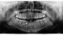
A healthy 14-year-old female presents to the office for a comprehensive exam prior to her getting braces. A panoramic radiograph revealed congenitally missing teeth (nos. 17 and 32). In between the lower one-third root apices of nos. 26 and 27, a circular radiopaque lesion was noted. It measured approximately 5 mm in diameter. Intraorally, the area was not tender to palpation, and both the patient and her parents were unaware of its presence.
You might also be interested in...
Oral pathology case: The common but not-so-common radiopacity
Pathology case: Two patients with the same diagnosis but different treatment
Diagnosis
Odontoma. An odontoma is a benign tumor containing a cluster of all the various component tissues of teeth.1 It compromises 67% of all odontogenic tumors.1 Odontomas will typically form in tandem with natural dentition. Most odontomas do not present with any signs or symptoms; however, depending on their location, they can hinder or delay the eruption of the permanent teeth.1 Odontomas occur more frequently in the maxilla, usually in the anterior part of the mouth, either over the crowns of unerupted teeth or between the roots of erupted ones.2 Lesions appear as a radiopaque mass, are usually well encapsulated, and can be easily removed without complication.1,2
Treatment
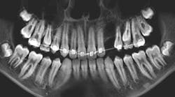
Since this patient was entering orthodontics, it was recommended that the odontoma be removed since it might be a hindrance to movement and/or have the potential to resorb the roots of the adjacent teeth. The tumor was removed without difficulty, and the patient commenced with orthodontics.
References
1. Goaz P, Wood N. Differential Diagnosis of Oral and Maxillofacial Lesions. Mosby; 1997:423-426. 2. Sapp JP, Eversole L, Wysocki G. Contemporary Oral and Maxillofacial Pathology. Mosby; 1997:147-149.


