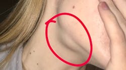
Definition and etiology
Sialolithiasis is the condition of salivary stone formation, causing inflammation and swelling of a salivary gland. It is most frequently manifested in the submandibular gland and Wharton’s duct papillae.1 The submandibular gland generally produces thicker, more mucoid saliva with a duct that can be easily infected with bacteria. The condition most commonly affects men 30–60 years old, with smokers having a higher risk for contraction.1
Related reading: The "non-bothersome" soft-tissue lesion
Calculi formation
Calculi or sialoliths form from a nidus of bacteria and grow with deposition of layers of inorganic substances (calcium carbonates and calcium phosphates). As the calculi grow, they block saliva from progressing down the duct and result in swelling of the salivary gland.
Early stage treatments generally involve the use of antibiotics as well as sialagogues, such as pilocarpine or even simple lemon drop candies. Advanced cases of sialolithiasis can be quite painful from both the swollen gland and the jagged-edged calculi trying to make their way down the duct. In those cases, surgical intervention may be required.
Case presentation and clinical exam
Patient history
Medical history was significant with the patient having cystic fibrosis (CF). In general, CF affects all of the patient's ductile systems by slowing motility and putting them at a higher risk for sialolithiasis. This was the patient’s third bout with similar, though less significant, symptoms.
Differential diagnostics
Differential diagnostic considerations included:
• Acute infection of the submandibular gland
• Neoplastic causes such as ductile papilloma, pleomorphic adenoma, or adenoid cystic carcinoma
• Autoimmune causes such as Sjögren’s syndrome or lupus
Treatment
Diagnosis and surgery
With the use of ultrasonography, Dr. Sniezek was able to confirm a diagnosis of sialolithiasis. The patient was scheduled for surgery using 1.3 mm diameter sialendoscopy.
Healing progressed well, though many more calculi worked their way down the healing duct over a three-week time frame. One-year follow-up shows a successful outcome with no recurrence of gland swelling and nicely healed, wider Wharton's duct papillae (figure 6).
Discussion
Sialolithiasis can be surprisingly painful for those with the condition. In the presented case, especially since the patient is also my daughter, I am very happy to report that the surgical intervention was a success!
The first few days postoperatively were miserable, and the unintended chemical burn on the end of her tongue (as seen in figure 5) made the pain even worse. Most cases can be resolved without surgery, but those that recur should be evaluated by a surgeon trained in sialendoscopy.
Reference
1. Marchal F, Dulguerov P. Sialolithiasis management: The state of the art. Arch Otolaryngol Head Neck Surg. 2003;129(9):951-956. doi:10.1001/archotol.129.9.951


