Adhesive endodontics: a whole new way to look at root canals
WRITTEN BY
Lori Trost, DMD, Editor
Traditionally, gutta percha has been the accepted standard for endodontic filling material because of its biocompatibility, thermoplastic nature, and retrievability. And, given its easy handling ability, it has been the “filler” material of choice for countless cases. However, gutta percha does possess one major disadvantage in that it does not seal or bond to the dentinal walls. In essence, it does not guarantee the absence of micro-leakage.
Much emphasis is now placed on the importance of the coronal seal mainly due to this focal point/entry of microorganisms into the canal and their ability to apically migrate. Several studies now indicate that coronal leakage is a critical indicator of the prognosis of root canal therapy1,2 Clinicians must take every step to ensure a seal is made in this key area as well as removal of any decay or bacteria.
Moving into the “adhesion” century, this drawback of gutta percha is now compensated for by the use of Resilon Products’ RealSeal™ (SybronEndo), Epiphany (Pentron), SimpliFill (LightSpeed USA), and InnoEndo (Heraeus Kulzer). Resilon Research LLC has created Resilon obturating points made of soft, thermoplastic resin that are radiopaque. Resilon has a unique property that distinguishes it from conventional gutta percha in that it forms a chemical bond to the cleared dentinal tubules, thus structurally creating a “monoblock” from dentinal tags, to sealer, to resin filler that extends the length of the canal. Each of these Resilon products comes with a resin sealer and a self-etching primer. And the beauty of Resilon is that although it mimics the handling properties of gutta percha, Resilon may be combined with all current obturation techniques - vertical compaction, cold lateral condensation, and a lateral/vertical combination, thus lessening the learning curve of a new product.
By the addition of two simple steps - clearing the smear layer and applying the primer - a clinician can achieve adhesion endodontics. The smear layer (deposits of organic and inorganic debris) created during the instrumentation process must be removed from the canal by the use of 17 percent EDTA and sodium hypochlorite. At the finish stage, either 17 percent EDTA or SmearClear, a 17 percent EDTA solution with surfactants (SybronEndo), can be delivered into the canal for one to two minutes to ready the canal for the primer stage. Care should be taken not to introduce sodium hypochlorite or absolute alcohol at this time because these materials will disrupt the resin sealer bond interface. Canals are then dried with paper points (with note taken that the canal wall(s) need not be completely dry because the resin sealer by nature is hydrophilic). Primer is then placed into the canals by a brush provided by the manufacturer or via a saturated paper point. Once again, care must be taken not to extrude the primer past the apical stop. Any excess primer should be removed via a paper point.
At this point, the obturation technique process follows the current choice of the clinician. The resin sealer is mixed and introduced into the canal. The Resilon master point is placed and the selected method of final obturation filling is performed.
The mindset to move into adhesion endodontics is truly simple. Virtually by the addition of two simple steps, a bit of time and a Resilon material, an adhesive seal that reduces microleakage can be achieved. This is truly a win for the patient and a good prognosis for long-term restorative care.
A case study
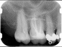
A 48-year-old woman presented with pain in her upper right quadrant. After taking her history and a radiograph, pulp testing confirmed irreversible pulpitis on Tooth No. 4 (Figure 1).
Figure 1
The patient was given a local anesthetic (Articaine 4 percent) and access was made into the pulp chamber. An initial file was positioned to the apex and a working length established via an apex locator (Elements Apex Locator, SybronEndo). An electric handpiece was set at a constant 350 rpm rotational speed. K-3 (SybronEndo) NiTi rotary files in their designated sequence were used to instrument the canals. Copius amounts of sodium hypochlorite irrigation was used during the instrumentation to clear debris from both canals.
Both canals were dried and RealSeal master points were fit with appropriate tug-back. Both master point cones were then placed in chlorhexidine until final placement. SmearClear was introduced into the canals via a syringe for two minutes. Paper points were used to dry the canals. Next, RealSeal primer was applied to the coronal portion of the canals using a brush. Saturated paper points were used to deliver the primer to the apical third of the canal. The goal was to saturate the canal walls evenly.
RealSeal resin sealer was extruded onto a small mixing pad and spatulated. A No. 25 file was coated with resin sealer and placed into each canal with a reverse spin to coat the walls. After that, the master cone was thinly coated with resin sealer and placed to the proper length.
Finally, a continuous wave of condensation was delivered using the System-B (SybronEndo). Excess master point was removed from the chamber with the heated tip. The preheated plugger stop was measured 3-4 mm of the prefitted measurement, then driven through the RealSeal, shy of the binding point. Apical pressure was maintained for a 10-second push and then the plugger was quickly withdrawn. To complete the obturation, the canals were back-filled with System-B. Note: The heat source was dialed down for the Resilon material to 160 degrees.
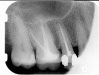
The postoperative radiograph can be seen in Figure 2. Subsequently, the patient proceeded with a build-up and crown. ■
References
1 Pisano D, DiFiore P, McClanahan S, Lautenschlager E, Duncan J. Intraorifice sealing of gutta-percha obturated root canal to prevent coronal microleakage. J Endod Oct 1998; 24(10):659-62.
2 Ray HA, Trope M. Periapical status of endodontically treated teeth in relation to the technical quality of the root filling and the coronal restoration. Int Endod J 1995; 28:12-18.
Lori Trost, DMD
is the managing editor of Woman Dentist Journal. She created the Center for Contemporary Dentistry in Columbia, Ill., in 1989. Her practice is known for being in the technological forefront. She is a member of the ADA and AGD.You may contact Dr. Trost at [email protected].


