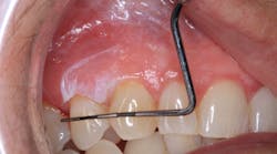
Dr. Stacey Simmons describes the oral pathology case of a patient who presented for her recare exam, unaware of a 9x6 mm lesion that was discovered during the clinical exam. Read more details about the case to see if you can diagnose this lesion.
Editor's note: This article first appeared in DE's Breakthrough Clinical with Stacey Simmons, DDS. Find out more about it and subscribe here.
A healthy 66-year-old female presents for her recare exam. Health history is noncontributory except for a hip replacement in the last year. Upon examination, a white, corrugated, irregular bordered lesion was noted on the upper right side on the attached tissue between teeth Nos. 5 and 6, measuring approximately 9x6 mm. The patient was unaware of its presence, and it was not tender to touch or palpation. Furthermore, the lesion was not able to be removed or scraped off.
What are your differentials, and how would you proceed with recommendations for this patient?
Send your answers to [email protected] or join our Facebook group to discuss this oral pathology case and more. Next month, we will discuss the final diagnosis and recommended treatment for this case.
For more pathology articles, click here.
Do you have an interesting oral pathology case you would like to share with Breakthrough’s readers? If so, submit a clinical radiograph or high-resolution photograph, a patient history, diagnosis, and treatment rendered to: [email protected]. We will let you know if we select your case.
Editor's note: This article first appeared in DE's Breakthrough Clinical with Stacey Simmons, DDS. Find out more about it and subscribe here.


