One of the most exciting discoveries for dentistry since the invention of the high-speed air turbine handpiece is the dental microscope. After 35 years of compromised positions at the dental chair while practicing as a hygienist and a dentist, my eyes and musculoskeletal system feel better now than 10 years ago. I'm enjoying dentistry more than ever since I graduated in 1979, and I'm providing better care to my patients. So what's my secret? It's U the dental microscope!
I've been using a dental microscope in every aspect of my practice for seven years. As I wheeled my first microscope on a cart from room to room, I thought I'd only use it for those hard-to-access posterior endo cases. But I soon realized how valuable it was for general dentistry. To see such beautiful margins for composites and amalgams was a thrill. To finally see upper second molars as clear as an anterior tooth was amazing! I soon learned that if I could see the problem, I could solve the problem. And the adventure began.
Not long after my first purchase of a dental microscope, it became very clear that one could not be shared easily between six rooms. Having to choose which patients would be treated with the microscope didn't seem fair — or ethical — because my results were so much better with the microscope.
In 1997 I purchased microscopes for the other five rooms, mounting each one on the rear wall of the operatory. Since there was no training manual for this new instrument, I taught my associates and hygienists how to operate efficiently with a microscope. By experimenting, note-taking, and documenting positions, we created new approaches to crown and cavity preps, and a pattern became evident for predictably using the microscope.
Dentistry became fun again, and my enthusiasm soared! The next step was to share my experiences with like-minded colleagues, Drs. Rick Schmidt and Glenn van As, who were using microscopes at that time for general dentistry. Since then, we have begun to share this new technology with other general dentists across the United States, teaching a grassroots, hands-on course in my office.
Many dentists ask me, "So, doctor, why use the dental microscope? I use loupes." I say, "I'm glad you are using magnification!" There is an old saying, "What you don't see, you don't know." Our ethical duty to practice with the very best diagnostics means that our patients deserve the best treatment we can provide. Microscopes allow me to practice the very best I can for every one of my patients. Since my visual IQ has increased with microscope use, I wouldn't practice dentistry without it! I love dentistry too much not to enjoy it every day. And I respect my patients too much not to do the best I can for them.
Visibility in the oral cavity has been one of the greatest challenges in practicing dentistry. Historically, dental professionals are taught to rely on tactile information when visibility is inadequate. But we can do better! In fact, we can see up to 24X power.
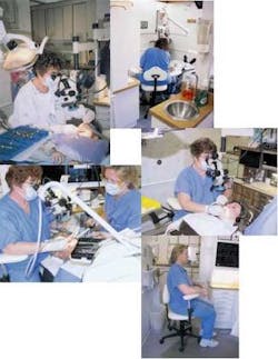
Clockwise, from far left ...
- Dr. Sheldon and a pedo patient
- Poor posture without microscope
- A comfort zone of total upper body support while working on a patient
- Surgical operating stool
- A comfort zone of total upper body support while working on a patient
The rationale for using dental microscopes is dependent on the magnification and illumination that allow us to practice better. While there is not general agreement about the exact magnification or illumination that is critical to practice optimum dentistry, the microscope can do better than the naked eye or loupes. Dr. Gary Carr, a colleague who uses microscopes, reported the resolution of the human eye, unaided by magnification, can distinguish two separate lines 0.2 mm apart.1 Anything less than 0.2 mm, the eye sees as one line. With 2X loupes, the improved resolution of the eye for the same two separate lines is 0.1 mm apart. With 4X loupes, the resolution is 0.05 mm. With 24X stereoscopic dental microscope use, the resolution becomes 0.0083 mm.2 This is superior to the tactile detection of an experienced dentist who, with a sharp explorer, can feel margins of 0.035 to 0.050 mm.
Illumination is another key factor in dental microscopes. While loupes allow up to 8X magnification, the lack of additional illumination limits their capability. The microscope overcomes this limitation with additional illumination provided by one of three different types of fiber-optic lights — halogen, metal halide, or xenon. As the magnification is increased, the light intensity remains very close to the same as with low power. Similar to sunlight shining through a magnifying glass, this light is better than any operatory light. I use my microscope light instead of the operatory light because it is brighter and more focused. I've found that it allows truer coloration for matching shades for composite colors and porcelain shades than is possible with any other light.
Besides superb magnification and illumination, the microscope allows me to operate in ergonomically correct positions. This technology creates a comfortable environment for me while working on patients. At the end of the day, I am ready for dancing lessons! Before microscopes, I was physically drained from having to contort myself on a daily basis. I believe microscopes can help prevent injury to spines — as well as necks, shoulders, and hands — due to poor posture in the dental chair.
The refined micro-motor skills required for microscope work are uniquely supported by a specially designed surgical operating stool. The microscope, along with the stool, keeps the operator in a comfortable, upright position with stereoscopic vision. The stool is fully adjustable for total support of the back, shoulders, and arms. You can be as relaxed as sitting at your desk reading a book.
The dental microscope has many uses besides endodontics. It is valuable for hard- and soft-tissue examinations, periodontics, amalgams, composites, crown and bridge, impressions, temporaries, cementing, pediatric dentistry, documentation, and patient education. For me, carving anatomy and finishing composites under the microscope are a joy as each impression is evaluated with the microscope. There is no guesswork on the margins.
Mehrabian stated that people understand 7 percent of what they are told and 55 percent from visual clues.3 In my practice, we have TV monitors connected to the microscopes, which are invaluable tools for patient education. Patients want to be involved in the decision-making process for their dental care. To be able to see the inside of the mouth on a TV screen is an extremely powerful patient motivator.
With all new technology there is a learning curve. Remember when computers were first being used? Even with the learning curve, computer technology was adopted almost universally because of its enhanced benefits for everyone. The dental microscope also requires a learning curve. That learning curve is followed by the benefits of saving our backs and producing the beautiful esthetic dentistry we want for our patients.
Diagnostic enhancing equipment has been used for surgical precision in medicine for years. Any woman who has undergone an ultrasound microscope for a cystic breast lesion has experienced this technology. The dental microscope allows a similar precision for our dental surgical procedures. This technology has been available since 1981, when Dr. Apotheker, a dentist, converted a medical operating microscope for use in endodontics. Then, in the early 1990s, Dr. Gary Carr partnered with Global Surgical Microscopes to design a surgical operating microscope and a dental operating stool for use in the dental operatory. Since presenting this at the 1992 meeting of the American Association of Endodontics, other dentists have become advocates of the dental microscope.
In November 2002, the Academy of Microscope Enhanced Dentistry was established by both specialists and general dentists. This group establishes policies and standards for teaching and practicing with dental microscopy. This wonderful new technology is here to stay in dentistry! Our dream is to see dental educators embrace this technology. We know that dental microscopes are key to assuring proper training of undergraduate students to provide the best dentistry in their practices.
References
- Carr GP, Hardin JF. Magnification and illumination in endodontics. Clarks Clinical Dentistry (New York, N.Y.; Mosby) 1998; 4:1-14.
- Van As GA. The use of extreme magnification in fixed prosthodontics. Dentistry Today June 2003; 22(6):93-99.
- Mehrabian A. Attitudes inferred from neutral verbal communications. Consult Psychology 1967; 31:414-417.
Doxey Sheldon, DDS
Dr. Sheldon has been in private practice in St. Louis, Mo., since 1979. She is a Fellow of the American College of Dentists and the International College of Dentists. In addition to lecturing, Dr. Sheldon teaches hands-on techniques for the dental microscope at her office.


