Aiming for success: radiologic techniques from analog to digital
By Marie George, RDH, MS
Success in dental radiography is achieved by obtaining a diagnostic image on the first exposure. A diagnostic image is one in which the image is the same size and shape as the object, has good detail, and has good density and contrast. These characteristics are critical to allowing the clinician to interpret the visual information on the image, accurately diagnose the situation, and appropriately plan treatment. Producing such a diagnostic image is influenced by exposure settings, exposure technique, and if using film, processing technique.
Success in dental radiography is achieved by obtaining a diagnostic image on the first exposure. A diagnostic image is one in which the image is the same size and shape as the object, has good detail, and has good density and contrast. These characteristics are critical to allowing the clinician to interpret the visual information on the image, accurately diagnose the situation, and appropriately plan treatment. Producing such a diagnostic image is influenced by exposure settings, exposure technique, and if using film, processing technique.
Exposure settings, including kvP, ma, and exposure time, influence the density and contrast of the image. As the kvP and ma settings are preset on contemporary radiographic equipment to produce a high quality beam of radiation with good penetrating ability, the exposure time is the only setting that remains variable and controllable by the radiographer, and should be adjusted accordingly as different areas of the dentition, with differing densities, are exposed.Exposure technique influences the remaining characteristics--size, shape and detail--of the image. Intraoral exposure techniques include paralleling and bisecting angle. Clinicians need to have a solid understanding of the principles of both paralleling and bisecting angle techniques, especially given the growing transition to digital radiography.
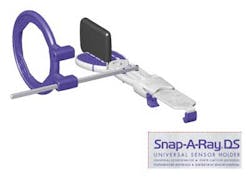
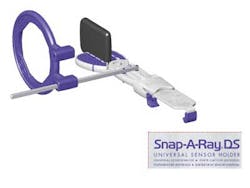
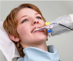
The basic principles of the paralleling technique are as follows:• The image receptor (film, PSP plate, or digital sensor) is placed at a distance to the tooth and parallel to the tooth in both the horizontal and vertical planes.• The central ray is aimed perpendicular to the tooth and image receptorThe parallel alignment of image receptor to tooth and perpendicular alignment of radiation beam to tooth/receptor minimize distortion; the placement of the receptor at a distance from the tooth and a long source-object distance provided by the PID minimize magnification. The result is an image that, without magnification or distortion, more accurately reflects the true clinical situation.The use of aiming devices (XCP instruments) with the paralleling technique further maximizes image quality by assisting the radiographer with correct vertical and horizontal alignment of the central ray, minimizing the chance for exposure errors, such as overlap, elongation, foreshortening, and cone cuts.While the paralleling technique, used in conjunction with aiming devices, is the preferred exposure technique to minimize errors and maximize image quality, the size and rigidity of direct digital sensors may necessitate use of the bisecting angle technique due to anatomical challenges or to avoid patient discomfort. The indication for using the bisecting angle technique is when anatomy prohibits parallel placement of the image receptor.The basic principles of the bisecting angle technique are as follows:• The image receptor is placed as close as possible to the tooth• The radiographer visualizes the angle formed between the image receptor and the long axis of the tooth, and then visualizes a line which bisects that angle• Central ray is aligned perpendicular to the visualized bisector lineBecause an aiming device is typically not utilized with the bisecting technique, the likelihood of exposure errors is significantly increased.
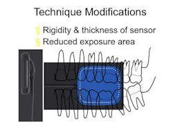
Modifications to exposure technique will be necessary when working with direct digital sensors due to the rigidity and thickness of the sensor, as well as the reduced imaging area. The rigidity of the sensor creates a challenge when placement of the sensor is limited by anatomy, such as a shallow or narrow palate, or the curvature of the bone in the area of the cuspids. This is further compounded by the reduced imaging area of the sensor. Where the flexibility of film or PSP plates conforms to the anatomy and allows for parallel placement, the rigidity of the sensor prohibits parallel alignment with the tooth. When the image receptor is not parallel to the tooth, the principles of the bisecting angle technique must be applied. Fortunately, new aiming devices that adjust for situations such as these are now available to assist the digital radiographer with correct alignment of the central ray, eliminating the guess work typically associated with bisecting angle technique.In summary, success in dental radiography is achieved when a diagnostic image is produced on the first exposure. Dental radiography is well in the midst of a digital transition. Digital radiography requires some modifications in technique due to the size and rigidity of the sensor and the reduced imaging area. Understanding the principles of both the paralleling and bisecting angle exposure techniques can assist the clinician in making appropriate technique modifications when necessary. The use of aiming devices and the paralleling technique, minimizes exposure errors and maximizes image quality, allowing you to be successful at analog or digital radiography in your practice.
Marie George, RDH, MS, is Curriculum Coordinator/Clinical Educator, MidAtlantic & TX, for DENTSPLY Professional.


