Editor's note: Originally published April 18, 2019. Updated March 2025.
Presentation
A 14-year-old male presented with a lesion of the anterior mandible first seen on periapical x-rays taken by his general dentist. The patient did not have symptoms and was unaware of any lesion prior to the x-rays.
Medical history and clinical exam
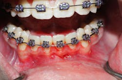
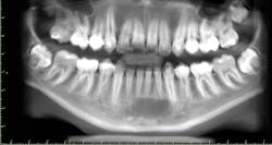
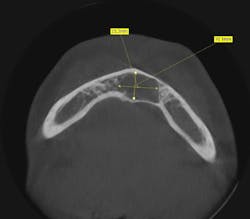
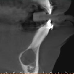
Clinically, there was no buccal or lingual expansion (figure 1). The patient’s medical history was noncontributory. A CT scan revealed a 1.5 cm x 1 cm well-defined, circular, hypodense area inferior to the lower incisors (figures 2–4).
Differential diagnosis
The differential diagnosis for a radiolucent lesion of the mandible in a 14-year-old includes:
- Keratogenic odontogenic tumor
- Simple bone cyst
- Dentigerous cyst
- Ameloblastoma
- Central giant cell lesion
Definitive diagnosis
Simple bone cyst
Discussion and treatment
A simple bone cyst is a lesion affecting males 60% of the time and found in the mandible more commonly than the maxilla. Most of these lesions are found in the 10- to 20-year-old age group. The hallmark of the diagnosis is a mostly empty lesion upon surgical entry.
Though the etiology is not fully understood, a theory involving past trauma has gained popularity, hence the lesion’s other name being a traumatic bone cyst. The lesion is also known as a solitary bone cyst. The theory postulates a traumatic incidence to the jaw that the patient usually doesn't remember, leading to an intrabony hematoma which then does not fully convert back to bone.
The radiographic appearance of a simple bone cyst can mimic many other odontogenic and nonodontogenic radiolucent lesions. Sometimes there is visible scalloping around teeth roots, and though usually unilocular with well-defined borders, these lesions can be multilocular with irregular borders.
A diagnosis is made only through surgical intervention. The cavity may be almost completely empty or contain some serosanguinous fluid. Very little soft tissue is obtained for histopathologic examination, which usually reveals fibrous connective tissue with some neural and vascular elements.
The treatment of simple bone cysts in the jaws is less aggressive than similar cysts found in long bones. In the jaw, curettage of the lesion is sufficient to trigger bony fill within six months. Long bone lesions may need intralesional steroids. It is important to submit even scant tissue found in the lesion, because rarely a lesion thought to be a simple bone cyst may have a keratogenic odontogenic tumor or ameloblastoma found on microscopic examination. Persistence or recurrence of the lesion is rare.
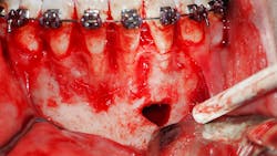
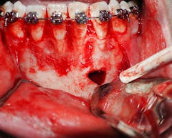
In this case, the patient underwent surgical exploration of the mandible. The lesion was mostly empty, though some tissue was submitted to pathology (figure 5). The tissue came back as fibrous connective tissue with some neural and vascular cells. The patient did well postoperatively.
Source
- Neville BW, Damm DD, Allen CM, Bouquot JE. Oral and Maxillofacial Pathology. 3rd ed. St. Louis, MO: Saunders; 2008:631-634.
About the Author

Ahmad Chaudhry, MD, DMD
Ahmad Chaudhry, MD, DMD, graduated summa cum laude from East Stroudsburg University of Pennsylvania. He then attended Harvard School of Dental Medicine, graduating with honors in 2000. He went on to earn his doctor of medicine degree from the State University of New York at Stony Brook in 2004 and completed his oral and maxillofacial surgery training in 2006, which included one year as a general surgery resident. Dr. Chaudhry practices at Lehigh Valley Oral Surgery and Implant Center in Bethlehem, Pennsylvania, and specializes in full-mouth rehabilitation with dental implants.
