Oral pathology cases can pop up in any dental specialty, so sharpening your detection skills is crucial. Learning from your colleagues is one of the best ways to improve clinical knowledge so you know how to diagnose and treat, and when to refer to a specialist.
Enjoy our roundup of pathology cases from DentistryIQ readers and keep these cases handy as a reference tool.
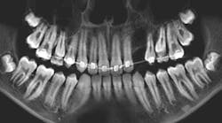
Patient: 14-year-old female entering orthodontics
- Congenitally missing teeth nos. 17 and 32
- 5 mm circular radiopaque lesion between the lower one-third root apices of nos. 26 and 27
The case of the ortho patient with a benign tumor
Patient: 38-year-old male
- 3 mm radiolucent/cystlike lesion surrounding a mesial/inferior-positioned no. 17
- 18: normal pocketing and normal testing to cold, percussion, and bite
A cystlike lesion that didn’t bother the patient
Patient: 36-year-old male
- 2x2 cm well-defined radiopaque lesion in the right submandibular region
- Lesion slightly palpable upon extraoral manipulation but not necessarily tender
Patients: 14-year-old female and 13-year-old male
Female—
- Large, multilocular radiolucent lesion within the entire ramus and posterior body of the right mandible
- 32 within the lesion
- Cortical expansion in the area
Male—
- Expanding radiolucency encompassing impacted tooth no. 11.
- Multilocular radiolucency extending from the maxillary midline to the area of the left permanent second molar and expanding into the left maxillary sinus
- Cortical expansion in the area
Two patients with the same diagnosis but different treatment
Patient: 54-year-old female
- Buccal mucosa bilaterally erythematous and atrophic
- Very tender to palpation
Manifestation of a common intraoral autoimmune inflammatory disease
Patient: 53-year-old male
- Incidental, well-defined, circular radiolucent area noted between roots of the mandibular right canine and first premolar
- Unremarkable health history
Incidental finding requires surgical excision
Have you recently encountered a pathology case in your practice that you’d like to share? Email your details to Bethany Montoya, BAS, RDH, the editorial director of Clinical Insights. We look forward to featuring you!
Editor’s note: This article first appeared in Clinical Insights newsletter, a publication of the Endeavor Business Media Dental Group. Read more articles and subscribe.
Vicki Cheeseman is an associate editor in Endeavor Business Media’s Dental Group. She edits for Dental Economics, RDH, Perio-Implant Advisory, DentistryIQ, and Clinical Insights.


