Editor's note: Originally published August 2016. Updated December 2023.
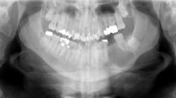
A healthy 66-year-old white male presented to the clinic as a new patient, having not seen a dentist in about six years. In the course of an otherwise normal cleaning, exam, and routine radiography, a large radiolucency was found in the left mid-body of the mandible. The patient denied any pain in the area, hadn’t noticed any new swelling, and denied paresthesia of his left inferior alveolar nerve (V3). His exam showed he was missing tooth no. 19. He explained that it was extracted by his previous dentist about seven years ago.
The rest of his dental exam was normal; there were no oral lesions or vestibular swelling. Extraorally there was no visual or palpable swelling in the area, and no cervical lymphadenopathy.
Differential diagnosis
- Residual cyst
- Keratocystic odontogenic tumor (KCOT)
- Ameloblastoma
- Traumatic bone cyst
- Calcifying epithelial odontogenic tumor (Pindborg tumor)
Final diagnosis: Residual cyst
Common things are common. The lesion the patient presented with should be highly suspicious for a residual cyst. A residual cyst is simply a periapical cyst that was not properly removed at the time the tooth was treated, and in this case during the extraction of tooth No. 19. The cyst remained in the mandible following the extraction, and for the following six years grew slowly and painlessly without the patient being aware of it.
Periapical cysts—also called radicular cysts—are easily the most common cysts of the jaws. Periapical cysts are inflammatory odontogenic cysts, which arise in response to the inflammation seen with necrosis and infection of the dental pulp. The cyst lining is derived from epithelial remnants of portions of the periodontal ligament (PDL). These cysts arise at the root apices of the offending tooth.
The inflammation resulting from the necrosis and/or infection of the dental pulp causes the formation of a granuloma at the root apex. Over time, this chronic inflammation stimulates the epithelial rests of Malassez in the periodontal ligament tissue to form the cystic lining. Then, through hydrostatic forces, the fluid in the cyst slowly causes the cyst to resorb bone and expand.
Treatment for the cyst outlined in this case first involved an incisional biopsy for definitive diagnosis of the lesion. Once the diagnosis of residual cyst was obtained, the patient underwent a second clinical procedure to remove the entirety of the cyst through an intraoral incision with enucleation and curettage. All walls of the mandible remained following removal; therefore, near-complete bony fill after healing should be seen. Recurrence of the lesion is extremely rare, so follow-up with normal dental visits and routine radiography should be sufficient.


