Let’s admit that the traditional methods of finding cavities — using our eyes, a radiograph, and the pick of an explorer — are a bit outdated. Our eyes can deceive us: is it a stained groove or actual decay? The shades of gray in a radiograph can lead to false positives, false negatives, and are only truly useful for interproximal decay. The explorer tip can be too large to fit into the full depths of fissures where decay lurks. Worse yet, a sharp explorer can break open a lesion that could have been conservatively remineralized.
RELATED READING | CAMBRA and caries detection technology
Fortunately, there are instruments that are far more accurate and sensitive for spotting carious lesions. Some of the technology has been around for decades and has been clinically proven to aid our diagnosis. The three basic camps use transillumination, fluorescence, or electrical resistance to detect differences in the microscopic structure of enamel and dentin. These differences may be displayed numerically, visually in colors, or audibly as beeps. Some systems offer software that can chart the recorded values for future comparison.
Transillumination
Fiberoptic Transillumination (FOTI) technology has been around for decades. It relies on the examiner to detect the subtleties in the transmission of light between healthy and decayed tooth structure and is still subject to operator error. Digital Imaging Fiberoptic Transillumination (DIFOTI) advanced this technique by allowing images to be captured and analyzed more objectively. Currently, the only product on the market that uses DIFOTI is CariVu.
Fluorescence
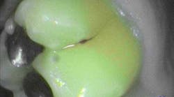
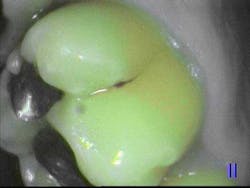
A light source, such as a laser or LED, emits light into the tooth. The tooth structures absorb this energy and then remit light, a phenomenon called fluorescence. The wavelength of the light that is emitted back from the tooth will vary according to its structure. Carious tooth structure is more fluorescent than healthy tooth structure, thus a device that can detect this difference gives us a noninvasive way of measuring the health of enamel and dentin.
Popular devices that utilize fluorescence include:
• The Spectra Caries Detection Aid — a 405-nm blue-violet LED light shows healthy tooth structure as green and carious areas in red. Image capturing software will also assign a number from 0 to 5 to aid diagnosis.
• DIAGNOdent — this 655-nm diode laser is calibrated to each patient as a baseline prior to use. Measurements are recorded as a two-digit number and audible beep.
RELATED READING |Are you sure I have a cavity?
• The Canary System — a 660-nm diode laser is used to collect both tooth fluorescence and photothermal radiometry (heat) readings. A proprietary software program combines the data of both reflected light and heat to give visual, numeric, and audible readings.
Electrical resistance
The Alternating Current Impedance Spectroscopy Technique (ACIST) detects lowered impedance (increased conductivity) in demineralized tooth structure compared with surrounding healthy tissue. Enamel and dentin that are carious are more porous. These pores contain ionic fluid that changes the ability of electrical current to flow through them. More modern devices will use variable frequencies for improved accuracy. The only product on the market that uses ACIST technology is the CarieScan PRO.
Advances in caries detection technology enable us to more accurately detect carious changes in tooth structure. Lesions that are questionable upon visual, tactile, and radiographic examination are better diagnosed using transillumination, fluorescence, and electrical resistance technologies. A spectrum of caries progression is more easily identified and thus a broader range of treatment options is available. The dental team can be more proactive in recommending preventive therapies or more confident in implementing surgical ones.


