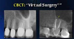
CBCT (cone beam computed tomography) uses systems that are ideal in capturing images of hard tissues especially in the maxillofacial region. One benefit of this technology is its ability to provide submillimeter resolution in terms of images. The images provided are also high in diagnostic quality. CBCT is now also used by a lot of dental professionals because of its short scanning period. It only takes about 10 to 70 seconds to complete scans.
RELATED |Guided implant surgery: Making sure dental implants are safe, predictable, and efficient
Radiation dosages were also reported as being 15 times lower than conventional CT scans. Increased use of this system can help dental clinicians in receiving imaging modalities that have the capacity of offering three-dimensional (3-D) representation of a patient's maxillofacial skeleton. CBCT performs this function with the least amount of distortion. This makes it a truly useful system for numerous professionals practicing dentistry.
A glimpse of what takes place during a CBCT session
In most cases, a single CBCT appointment or session involves a variety of procedures that can be completed in less than one hour. These include registration, scanning, positioning, reconstruction, and verification of the scan. I personally advise my clients to visit my clinic 30 minutes before their appointment to handle the preparation process efficiently. This can also give me enough time to assess their current dental and oral condition.
I also personally use CBCT technology in the event that three-dimensional systems are needed for the correct diagnosis, planning of treatment, and management of the present condition of the maxillofacial complex and jaws. In my years of dental practice, I recognized how useful CBCT is in improving diagnostic and treatment planning particularly in the following instances and cases.
For dental implants
CBCT works in immediately spotting or locating anatomic structures, incisive canal, submandibular fossa, mandibular canal, and maxillary sinus. It is also helpful in the correct assessment of the shape and size of the ridge as well as the quality and quantity of bones. It is valuable in assessing the required number of implants identifying the need for a sinus lift or bone graft, and determining the importance of using implant planning software.
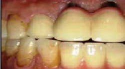
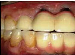
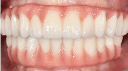
Here is a clinical photograph showing multiple implants applied five years ago.
When using CBCT, the amount of bone loss will be clearly detected as well as the damages in one's teeth and how dental implants can help.
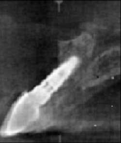
This is a picture taken from CBCT that clearly shows complete buccal plate destruction.
This is another CBCT image that has helped me assess bone density during the course of treatment.
For maxillofacial and oral surgery
CBCT is useful in spotting the relationship between mandibular canal and third molar roots. It also helps in foreign objects and impacted teeth localization, orthognathic surgery planning, and facial fractures and asymmetry assessment. I have also noticed the usefulness of CBCT in maxillofacial and oral pathology. It is valuable in localizing and characterizing lesions found in the jaw. It also evaluates how lesions affect the jaw in third dimensions especially those effects related to bilateral symmetry, cortical erosion, and expansion. It also evaluates how lesion relates to one's teeth and some of its other most vital structures.
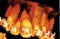
This figure shows how effective CBCT system is in clearly showing more lesions than other software that capture images.
For orthodontics treatment and TMJ
CBCT is useful in planning orthodontic treatments for complex cases. This holds true if the three-dimensional information is required as a means of supplementing or substituting other forms of imaging. I also use CBCT for patients who have cleft palate, root resorption and angulation, and impacted tooth. CBCT systems are also useful in dealing with temporomandibular joint (TMJ). The systems are effective in the accurate assessment of the TMJ's structures, thereby guaranteeing its correct diagnosis and treatment.
Benefits of using CBCT systems
Using CBCT systems in my dental practice has truly helped me ensure that my patients receive the best out of the services I provide to them. One of the benefits of CBCT is low radiation doses compared to medical CT. It also adds a level of comfort for patients. It makes them feel at ease because it is conducted in an open environment in a relaxed seating position while also facing out.
The scan takes about 20 seconds and allows the immediate availability of images on screen. These images can be imported into another software. All the benefits provided by CBCT, especially in achieving safe and reliable results when performing a number of dental procedures, makes it a truly valuable tool in my long years of practice in the field of dentistry.
ADDITIONAL READING ...
Implants made easy
"Mission Critical" technologies
Dr. Patrick Crawford is owner of Pat Crawford Dentistry. He enjoys writing articles that help people understand the importance of dentistry and maintaining a healthy mouth. He practices dentistry in Kenosha, Wisconsin.
