Head and neck cancer, including oral cancer, cancer of the larynx, and cancer of the pharynx, is the sixth most common type of cancer, with approximately 643,000 worldwide cases reported annually.(1)
About 40% of tumors diagnosed in the head and neck area are diagnosed as squamous-cell carcinoma.(2) The five (5) year survival rate for these kinds of oral cancer ranges from 81% (patients with localized disease), 42% (with regional disease), and 17% (patients with metastasis.(3) This rate has remained very high and relatively constant in recent decades.(2)
The treatment of oral cancer often produces major changes in speech, chewing, swallowing and oral health, which in addition to the disease per se, affects the social life and self-esteem of the affected person.(4,5,6)
Unlike other areas of dentistry, where new methods allow early diagnosis of many diseases, the oral examination has not changed much in the last decades. Currently, we already have some chairside additional tests available as adjuncts to the oral examination. This article will focus on light-based detection systems and fluorescence visualization.
Light-based detection systems use chemiluminescence, blue-white LED, and autofluorescence as light sources. Three products, ViziLite Plus (Zila Pharmaceuticals), Orascoptic DK (Sybron Dental), and Microlux/DL (AdDent Incorporated), use light-based detection. ViziLite Plus uses a chemiluminescent light stick.
Orascoptic DK and Microlux/DL use a blue-white LED fiberoptic light. Another light-based detection technology is fluorescence visualization, and that is the mechanism of action of VELscope. Unlike the other light-based systems, the fluorescence does not require a pre-rinse.(7,8)
The VELscope technology stimulates epithelial cells and stroma by a blue light (400-460 nm). It is the self-fluorescence of the tissues that allows detection of changes morphology and composition of the tissues in a non-evasive manner. However, this additional examination does not replace the oral examination and scalpel biopsy, which remains the golden standard of diagnosis.(8)
We are using the VELscope in our multidisciplinary clinic as an oral cancer-screening device. It does not diagnose cancer, but is an adjunct to the oral examination. It is, however, according with our experience, a fantastic non-invasive screening device. It is a misconception that some dentists or dental hygienists hold that this device will determine whether or not the lesion is cancerous. It does not. It simply aids in finding abnormalities not visible to the naked eye.
We personally do not charge a separate fee for the VELscope because we do not want cost to enter the patient’s decision as to whether or not to have the exam. We feel that our practice should provide the highest level of care, incorporating the latest technology in detection and treatment.
The JADA study(7) states that VELscope does not find 50% more abnormalities than doing a good visual exam with a regular white light. In fact, it is our belief that this is not a problem. If we send a few extra people to the oral surgeon, and the biopsy is negative, we feel better than if we missed a cancerous lesion.
What disturbs us is if we tell the patients that they are okay, and something is missed because they did not have a biopsy. This might happen in 5 out of 10 of the patients, and we feel it is unacceptable.
The subject of oral cancer detection is an exciting and constantly progressing area of research and technology. Detection tools are becoming increasingly accurate and less invasive as studies continue to be published in order to determine the sensitivity and specificity of each detection mechanism. More studies and education materials, as well as supporting information on how they can be best utilized in our practice, are essential for success.
As healthcare providers we play a vital role in patients’ oral and overall health. Integration of the adjuncts discussed here can help detect hidden lesions before they have the chance to progress, and this will improve patients’ chances of living a healthy life. The detection of this disease in its early stages constitutes an important facet of prevention and is the key to survival.(8)
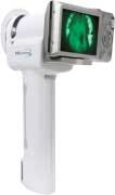
The last Velscope model with an intraoral camera incorporated
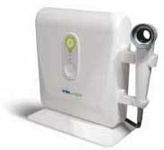
Our model
References 1. Fedele S. Diagnostic aids in the screening of oral cancer. Head & Neck Oncology 2009. 2. Epstein JB, Silverman S Jr, Epstein JD, Lonky SA, Bride MA. Analysis of oral lesion biopsies identified and evaluated by visual examination, chemiluminescence and toluidine blue. Oral Oncol. 2008 Jun; 44(6). 3. Patton LL, Epstein JB, Kerr AR. Adjunctive techniques for oral cancer examination and lesion diagnosis: a systematic review of the literature. J Am Dent Assoc. 2008 Jul;139(7).


